1.1 Introduction
A bone is an organ, not just a hard material. It contains mineralized bone tissue, marrow, blood vessels, nerves, and two covering membranes: the periosteum outside and the endosteum inside. Where it forms a joint, it is covered by cartilage.
This matters to the reconstructive surgeon for one reason. A piece of bone can keep its shape after all its cells have died. A dead segment and a living segment can look the same on X-ray. Whether bone is alive depends on its cells, its membranes and its blood supply, not on its shape.
This chapter describes the parts of a bone that can be seen with the naked eye: the shapes of bones, the regions of a long bone, the covering membranes and the marrow, and the flat and facial bones that plastic surgeons operate on most.
Flowchart: Overview of the macroscopic anatomy of bone
- A bone (the organ)
- Shape
- Long, short, flat, irregular, sesamoid
- Regions of a long bone
- Epiphysis, physis, metaphysis, diaphysis
- Coverings and contents
- Periosteum, endosteum, marrow, cartilage
- Flat and facial bones
- Calvarium, maxilla, mandible, orbittables and diploë; buttresses; sinuses
- Shape
1.2 Functions of bone
Bone has mechanical functions and metabolic functions.
| Function | What it means | Example in reconstruction |
|---|---|---|
| Support | Gives the body its frame and its shape | The mandible and the zygoma give the lower face and the cheek their projection |
| Protection | Covers the brain, the eye and the chest organs | A cranial vault defect must be closed to protect the brain; the orbital floor supports the eye |
| Movement | Acts as a lever for muscles, through joints | The mandible moves at the temporomandibular joint for chewing and speech |
| Mineral store | Holds about 99% of the body's calcium and most of its phosphate | Bone is removed to keep blood calcium normal; this can thin the skeleton in disease |
| Blood formation | Red marrow makes blood cells | Cancellous bone from the iliac crest carries marrow cells with it |
| Hormone production | Bone cells make FGF23, a hormone that controls phosphate loss by the kidney (Shimada 2004) | Explains some rare diseases of poor mineralization |
1.3 Classification of bones by shape
Bones are first described by their shape.
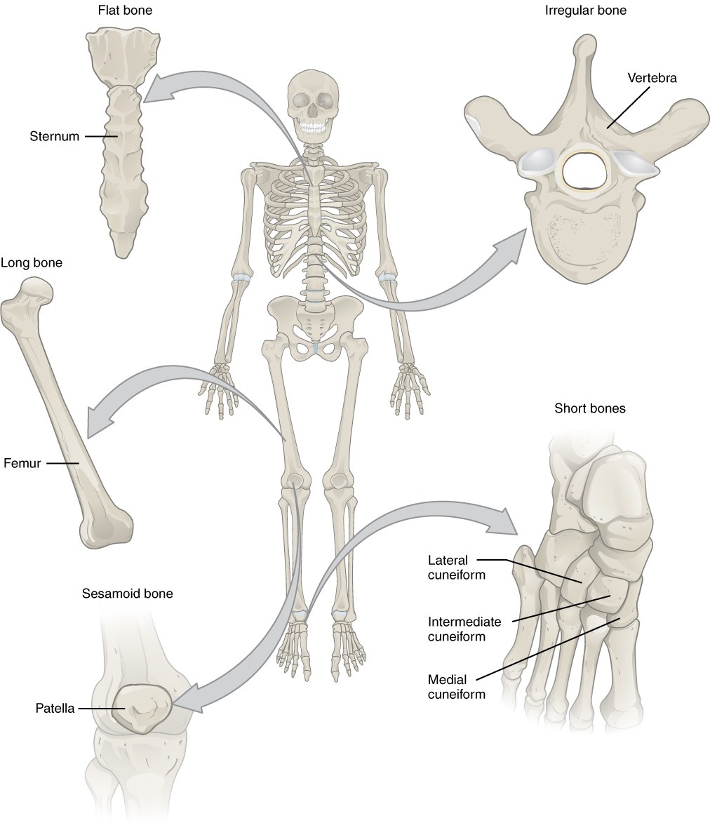
| Shape | Description | Examples |
|---|---|---|
| Long | A tubular shaft with two expanded ends. It is "long" because of its shape, not its size: a phalanx is a long bone. | Femur, tibia, fibula, radius, metacarpals, phalanges |
| Short | Roughly cube-shaped. A thin cortex around a cancellous center. Several joint surfaces. | Carpal and tarsal bones |
| Flat | Thin plates. Protect organs and give a large surface for muscle attachment. | Skull vault, sternum, scapula, ribs |
| Irregular | Complex shapes that do not fit the other groups | Vertebrae, most facial bones (maxilla, ethmoid, sphenoid) |
| Sesamoid | Develop inside a tendon; change the direction of pull of the tendon | Patella, sesamoids of the thumb |
Shape is only one way to describe a bone. Three other descriptions are used in the following chapters, and they answer different questions:
- Architecture: is the bone dense (cortical) or a network of struts (cancellous)?
- Matrix organization: is the collagen arranged at random (woven bone) or in ordered layers (lamellar bone)?
- Development: did the bone form directly from membrane (intramembranous) or through a cartilage model (endochondral)?
For example, the shaft of an adult fibula is long by shape, mainly cortical by architecture, lamellar by matrix and endochondral by development. The parietal bone is flat, has two cortical tables with cancellous bone between them, is lamellar, and is intramembranous.
1.4 Regions of a long bone
A long bone has four regions, from the end to the middle.
Flowchart: Regions of a long bone, from the end to the middle
- Epiphysisthe end; covered by joint cartilage
- Physis (growth plate)cartilage; adds length in children
- Metaphysisthe flared part; thin cortex, much cancellous bone
- Diaphysisthe shaft; thick cortical tube around the marrow cavity
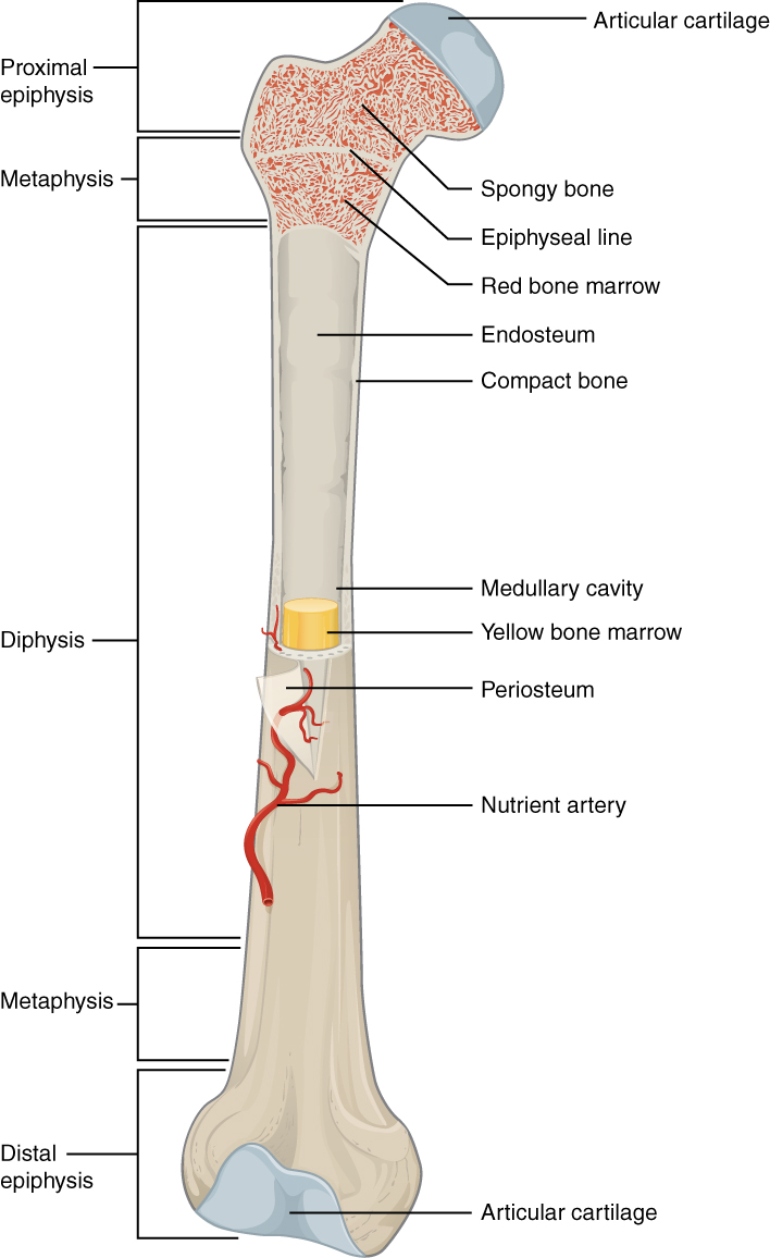
| Region | Structure | What it means for the surgeon |
|---|---|---|
| Diaphysis | A tube of thick cortical bone around the marrow (medullary) cavity. A tube resists bending and twisting with little weight. | The fibula flap is taken from the diaphysis. A diaphyseal defect must be restored for length, alignment and rotation. |
| Metaphysis | Thin cortex, many trabeculae, rich blood supply | Heals quickly. Is a source of cancellous graft (for example the distal radius and the proximal tibia). |
| Physis | A plate of cartilage between the epiphysis and the metaphysis in a growing child | Injury can stop growth. A fibula can be transferred with its physis to keep growth in a child. |
| Epiphysis | Cancellous bone under a dense subchondral plate and joint cartilage | The joint surface must be restored and supported, not only united. |
1.5 Periosteum, endosteum and marrow
1.5.1 Periosteum
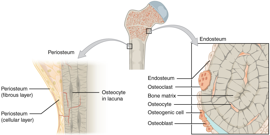
The periosteum covers the outer surface of almost every bone, except the joint surfaces. It has two layers:
- An outer fibrous layer of dense collagen with vessels and nerves. Tendons and ligaments blend into it.
- An inner cambium layer, next to the bone. It contains osteoprogenitor cells: cells that can become bone-forming cells (osteoblasts). These cells make new bone during growth and after a fracture.
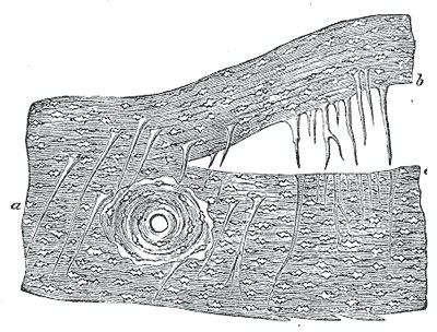
The periosteum is fixed to the bone by bundles of collagen called Sharpey fibers, which run from the fibrous layer into the cortex.
The periosteum changes with age:
- In children it is thick, loosely attached and very active. A child's fracture forms large callus, and a child's periosteum can make new bone even after the bone underneath is lost.
- In adults it is thin and more firmly attached, and its cambium layer has fewer cells (Allen 2004).
The periosteum has three jobs that matter in surgery. It supplies bone-forming cells; in mice, a specific stem cell in the periosteum forms the bone made directly from membrane, without cartilage (Debnath 2018). It carries blood vessels into the outer cortex. It is the layer through which muscle and tendon attach. Every time we strip periosteum to expose a fracture or to place a plate, we remove cells and blood supply together.
1.5.2 Endosteum
The endosteum is a thin layer of cells that lines all the inner surfaces of bone: the marrow cavity, the surfaces of the trabeculae and the vascular canals of the cortex. It also contains osteoprogenitor cells. The total inner surface is much larger than the outer surface, so much of bone formation and removal happens on the endosteal side.
1.5.3 Marrow
- Red marrow makes blood cells. It also contains stromal cells, which support blood formation and include skeletal stem cells.
- Yellow marrow is mainly fat.
At birth all marrow is red. With age, red marrow in the limbs is replaced by yellow marrow. In adults, red marrow remains mainly in the pelvis, vertebrae, sternum, ribs, skull and the upper ends of the femur and humerus. This is one reason why the iliac crest is the usual source of cancellous bone graft: its marrow still contains many cells.
1.6 Joint cartilage and subchondral bone
Joint (articular) cartilage is hyaline cartilage. It has no blood vessels and no periosteum, so it has almost no ability to repair itself. Under it lies the subchondral plate, a layer of dense bone that passes joint forces into the cancellous bone below.
In the face, the temporomandibular joint follows the same rule. A condylar fracture can unite and still leave the joint not working properly if the condyle's shape or position is lost.
1.7 Flat bones: the calvarium
The skull vault (calvarium) is the most typical flat bone. It has three layers:
- An outer table of cortical bone, covered by the pericranium (the periosteum of the skull).
- The diploë, a layer of cancellous bone with marrow and large valveless veins (diploic veins).
- An inner table of cortical bone, against the dura.
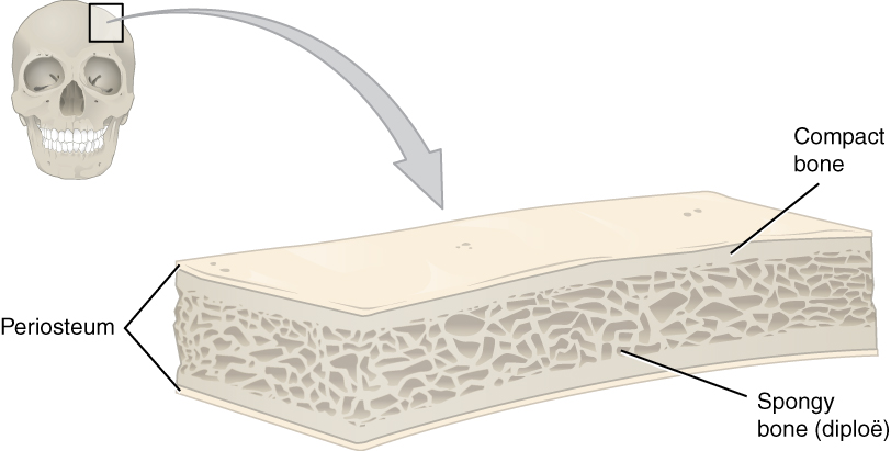
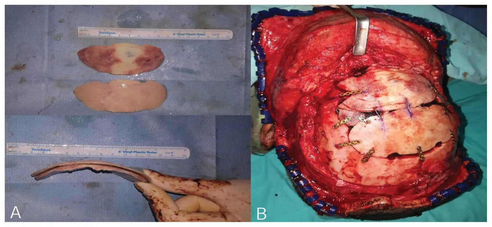
The diploë is the plane used to split the skull. A split calvarial graft is taken by separating the outer table from the inner table through the diploë, leaving the inner table to protect the brain. Three anatomical facts guide this:
- Thickness varies with the site, the person's sex, race and weight. In 200 adult cadavers, Pensler and McCarthy (1985) measured these differences at four common donor points. The parietal bone is the usual donor site because it is thick and lies away from the frontal sinus and the superior sagittal sinus.
- The diploë is absent in infants. It forms during childhood, so the skull of a young child cannot be split.
- The calvarium is dense and membranous in origin. Calvarial graft keeps its volume better than iliac crest graft when placed on the face. This was first explained by its membranous origin, and later mainly by its dense cortical structure.
Not all flat bones are built like the calvarium. The sternum is a shell of cortex around marrow-filled cancellous bone. The scapula is a thin plate with thick borders, which is why a scapular flap is taken from the lateral border. The term "outer table, diploë, inner table" applies only to the skull.
1.8 The facial skeleton
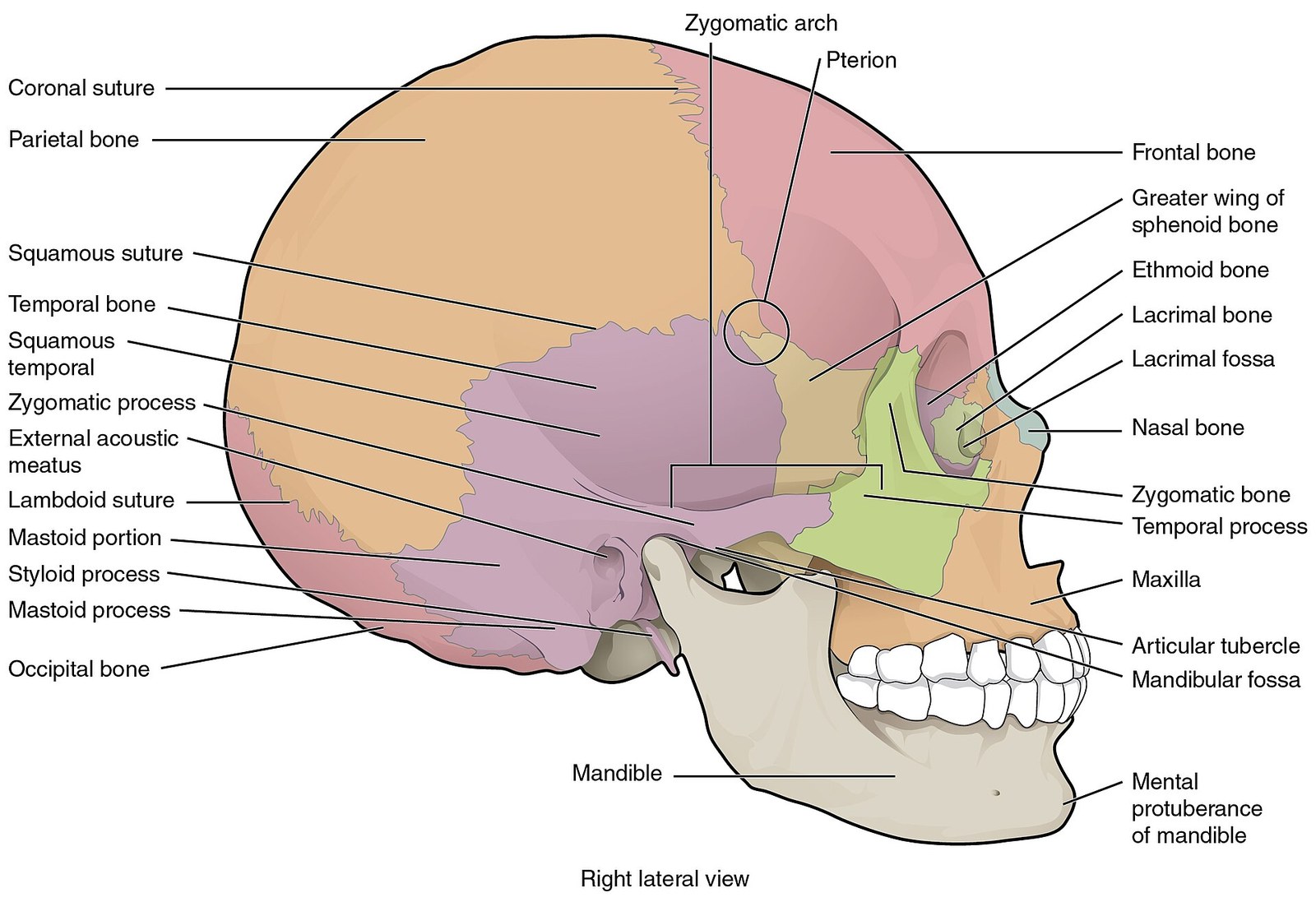
Most facial bones are classed as irregular. Their shape is complex, many are thin, and several are hollow.
1.8.1 Thin bone, thick pillars: the buttresses
The midface is mostly thin bone around the sinuses and the orbits. Its strength comes from thicker columns and bars of bone called buttresses. They carry the force of chewing up to the skull base.
| Direction | Buttress |
|---|---|
| Vertical | Nasomaxillary (medial): along the edge of the nose to the frontal bone Zygomaticomaxillary (lateral): from the molar region through the zygoma to the frontal bone Pterygomaxillary (posterior): behind the maxilla to the skull base |
| Horizontal | Frontal bar; infraorbital rims and zygomatic arches; the palate and alveolus; the mandible |
Midface fractures are reduced and fixed at the buttresses, because that is where the bone is thick enough to hold screws and where load passes. Missing buttress can be replaced with bone graft at the first operation (Gruss and Mackinnon 1986).
1.8.2 Sinuses
The maxillary, frontal, ethmoid and sphenoid bones contain air spaces lined by mucosa (pneumatized bone). A defect that opens into a sinus or the mouth is exposed to bacteria. A bone graft placed there needs good soft tissue cover and a seal between the cavity and the graft.
1.8.3 The orbit
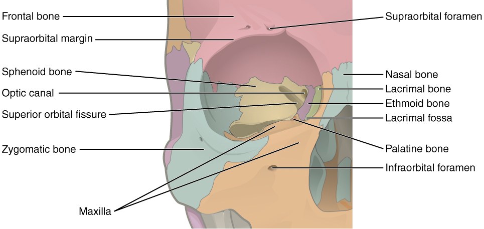
Seven bones form the orbit: frontal, zygomatic, maxilla, lacrimal, ethmoid, sphenoid and palatine. The orbital floor and the medial wall are paper-thin and break easily. Reconstruction of the orbit must restore its shape and volume, because a small change in volume moves the eye.
1.8.4 The mandible
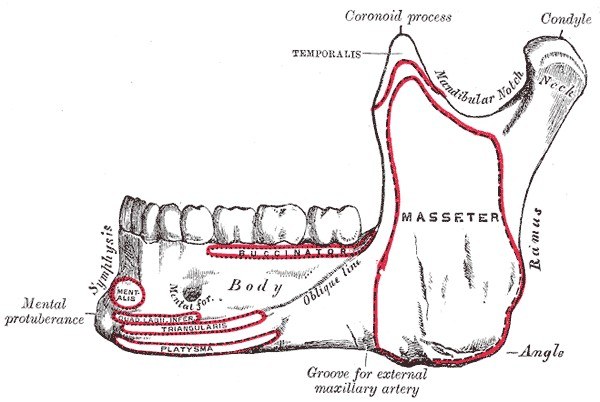
The mandible behaves more like a long bone than the rest of the face. It is a U-shaped arch of thick cortical bone, with a canal for the inferior alveolar nerve and vessels, and a joint at each end. It is loaded every time the patient chews. When a segment is lost, the remaining parts are pulled out of place by the muscles attached to them.
1.8.5 Region and reconstructive need
| Region | Main anatomical feature | What the reconstruction must give |
|---|---|---|
| Calvarium | Two tables and diploë; dura beneath | Protection and contour |
| Orbit | Thin walls; fixed volume | Shape and volume, to keep the eye in position |
| Midface | Thin bone, buttresses, sinuses | Restored buttresses, projection, separation of mouth and nose |
| Mandible | Loaded arch of cortical bone with two joints | Continuity, occlusion, bone height for teeth |
| Long bone shaft | Cortical tube | Length, alignment, rotation and strength |
| Growing bone | Physis, sutures | Union now, and growth later |
1.9 Short, irregular and sesamoid bones
Short bones have a thin cortex around cancellous bone, and much of their surface is covered by joint cartilage. Vessels can enter only through the small areas not covered by cartilage. The scaphoid is the best example: its main vessels enter at the distal end, so after a waist fracture the proximal pole can lose its blood supply.
A sesamoid bone works with its tendon. The patella increases the leverage of the quadriceps. Its repair must restore the tendon mechanism, not only the bone.
1.10 Summary
- A bone is an organ. Its shape can survive after its cells have died.
- Bones are described by shape, architecture, matrix organization and development. These describe different things.
- A long bone has an epiphysis, physis, metaphysis and diaphysis. The diaphysis is a cortical tube; the metaphysis is mostly cancellous.
- The periosteum has a fibrous layer and a cambium layer of bone-forming cells. It is thick and active in children. Stripping it removes cells and blood supply.
- The calvarium has an outer table, diploë and inner table. A split graft is taken through the diploë.
- The midface is thin bone held by buttresses. The mandible is a loaded cortical arch.
References
- Shimada T, Hasegawa H, Yamazaki Y, Muto T, Hino R, Takeuchi Y, et al. FGF-23 is a potent regulator of vitamin D metabolism and phosphate homeostasis. J Bone Miner Res 2004;19:429–435.
- Allen MR, Hock JM, Burr DB. Periosteum: biology, regulation, and response to osteoporosis therapies. Bone 2004;35:1003–1012.
- Debnath S, Yallowitz AR, McCormick J, Lalani S, Zhang T, Xu R, et al. Discovery of a periosteal stem cell mediating intramembranous bone formation. Nature 2018;562:133–139.
- Pensler J, McCarthy JG. The calvarial donor site: an anatomic study in cadavers. Plast Reconstr Surg 1985;75:648–651.
- Gruss JS, Mackinnon SE. Complex maxillary fractures: role of buttress reconstruction and immediate bone grafts. Plast Reconstr Surg 1986;78:9–22.