7A.1 Introduction
Every method we use today was the answer to a problem that the previous method could not solve, and each answer created a new problem of its own. Splints could not hold a reduction, so casts were invented. Casts left joints stiff, so surgeons fixed the bone. Rigid fixation damaged the blood supply, so fixation became more biological. Grafts could not survive in a poor bed, so bone was moved with its own vessels.
In this chapter each step is told in the same three parts: the problem, what changed, and what was still missing. Read in order, the missing part of one step becomes the problem of the next.
Flowchart: Five eras of fracture care and bone reconstruction
- Immobilize and observeantiquity to the 1800s
- Casts, asepsis, X-rays, first implants1850s to the 1900s
- Rigid fixation or functional treatment1925 to the 1960s
- Biology, regeneration, reconstruction1968 to today
Running alongside: the facial skeletonfrom Hansmann's mandible plates (1886) to virtual planning
7A.2 Immobilize and observe (Antiquity to the 1800s)
For most of history, treating a fracture meant holding the limb still and waiting. The tools were splints, bandages and traction, and the biology of healing was a mystery.
7A.2.1 Splints and bandages in the ancient world (c. 1600 BCE)
The problem. A broken limb had to be held still, and nobody could see the bone.
What changed. The Edwin Smith papyrus, a c. 1600 BCE copy of a much older text and the oldest known trauma treatise, describes bandaging and splinting and advises immobilization for fractures. Egyptian splints were bark wrapped in linen. Hippocrates described wooden splints and reduction, and advised exercise to prevent muscle wasting during immobilization.
What was still missing. Splints slip and bandages slacken. Bed rest and restricted activity remained the standard treatment for centuries.

7A.2.2 Stiffened bandages (AD 30–1500s)
The problem. Splints and soft bandages could not keep a reduced fracture in position.
What changed. Celsus (AD 30) described bandages stiffened with starch. Arab physicians used lime and egg white, the school of Salerno flour and egg, and medieval bonesetters egg white, flour and fat. In the 16th century Ambroise Paré made casts of wax, cardboard, cloth and parchment that hardened as they dried.
What was still missing. They were slow to prepare and to harden, and bed rest remained the standard.

7A.2.3 Where does new bone come from? (1700s–1867)
The problem. Splinting held the bone, but nobody knew what made it heal, or whether transplanted bone stayed alive.
What changed. In the 18th century Duhamel described an osteogenic layer of the periosteum, the "cambium layer". Ollier's two-volume experimental study of 1867 showed that transplanted periosteum and bone could survive and produce bone.
What was still missing. Von Haller, and later Macewen, held that the periosteum only guides growth. Whether a graft is a passive scaffold or an active source of bone stayed disputed for a century, and it still underlies how grafts are classified.
7A.3 Casts, asepsis, imaging and the first implants (1850s to the 1900s)
By the mid-nineteenth century immobilization was the standard, and its limits were clear. Three developments changed the field: a cast that could be molded and worn, an operation that patients could survive, and a way to see the bone. Together they made the first implants possible.
7A.3.1 Plaster of Paris (1851)
The problem. Stiffened bandages hardened slowly, and bed rest remained the rule.
What changed. Military surgeons Larrey, Seutin, Mathijsen and Pirogov developed stiff dressings. In 1851 Antonius Mathijsen, working in Haarlem, applied plaster of Paris bandages, which set quickly and could be molded to the limb. Ambulatory treatment of fractures followed directly.
What was still missing. Plaster is heavy and tolerates water poorly (fiberglass casts appeared in the 1970s), and prolonged immobilization stiffens the joints it crosses.
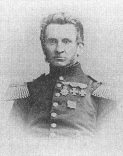
7A.3.2 Anesthesia and antisepsis: opening a fracture becomes survivable (1860s–1880s)
The problem. Opening a fracture usually meant infection, sometimes death, so bones were treated closed, at the price of deformity and stiff joints.
What changed. Anesthesia and Lister's antiseptic method made surgery tolerable and, for the first time, survivable. Iron wire had been reported for fractures as early as 1775, and cerclage wiring now became practical.
What was still missing. Wire held fragments poorly. Fixation was unstable, casts were still needed, and early reports describe rare successes and frequent catastrophes.
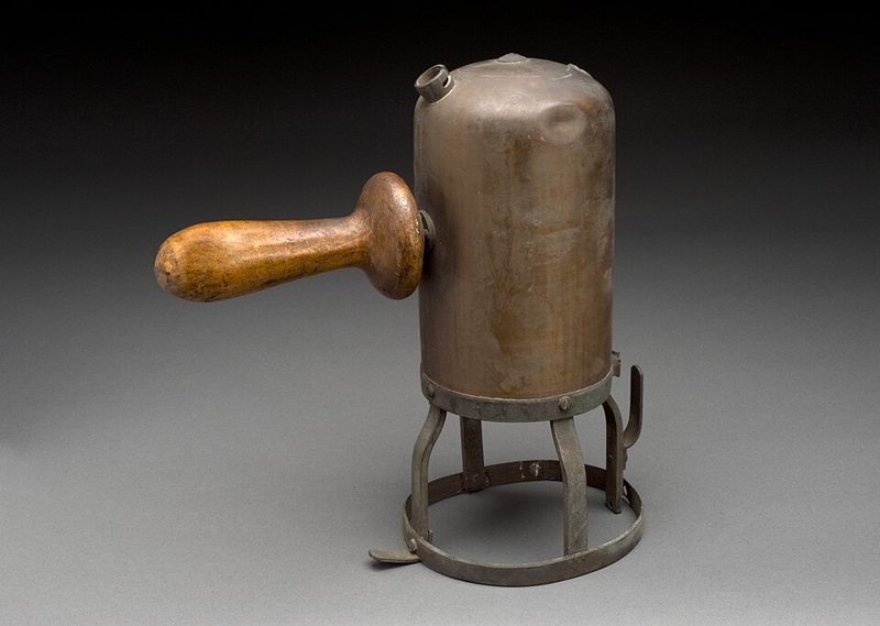
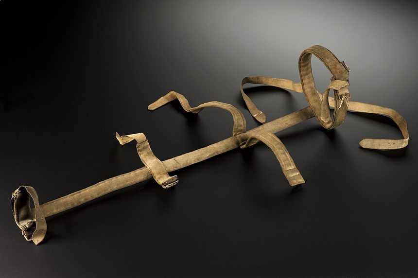
7A.3.3 Traction and the Thomas splint (1875–1918)
The problem. Femoral shaft fractures, especially open ones, carried a very high mortality, and healing meant long inactivity.
What changed. Hugh Owen Thomas, from a family of bonesetters, described his hip and knee splints in 1875. Used for femoral fractures in the First World War, the Thomas splint was credited with cutting mortality from open femur fractures from about 80% to 20%.
What was still missing. Survival improved, but treatment still meant weeks in traction, and the bone was held only indirectly.
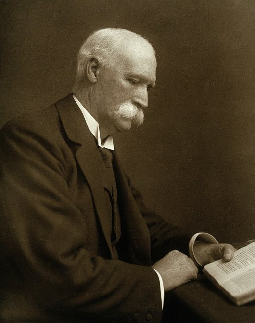
7A.3.4 The first bone allograft (1878–1881)
The problem. A large segment of bone lost to disease could not be replaced.
What changed. The Scottish surgeon William Macewen reconstructed a child's humerus with donor bone in 1878–79 and reported it in 1881: the first inter-human bone allograft. Anesthesia and antisepsis had made it possible.
What was still missing. Nobody could say whether the graft lived or merely served as a scaffold, the unresolved question of Duhamel and Ollier. Allograft use later waned, and banked bone arrived only after the Second World War.
7A.3.5 The first plates and screws (1886–1912)
The problem. Wire and plaster could not hold fragments in position, and antisepsis had at last made an implant survivable.
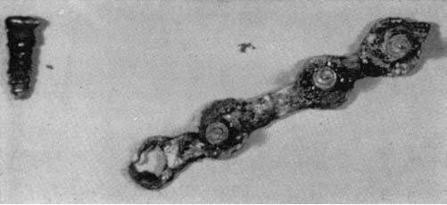
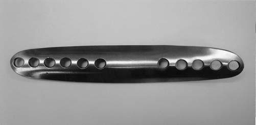
What changed. Carl Hansmann (Hamburg, 1886, including two mandible fractures), Arbuthnot Lane in the 1890s and Albin Lambotte, who coined the word "osteosynthesis" in 1904, screwed metal plates to bone. Sherman followed in 1912.
What was still missing. Early steel corroded, and Lane's plate was abandoned. Better alloys cured the corrosion, but the thin plates of Lambotte and Sherman were too weak. The fear of infection remained.
7A.3.6 Wolff: bone adapts to load (1892)
The problem. Why does bone change its architecture after injury and with use?
What changed. Julius Wolff's Das Gesetz der Transformation der Knochen (1892) proposed that healthy bone adapts to the loads placed on it.
What was still missing. The law describes an outcome, not the cellular sensor or a safe loading dose, and it cannot be transferred to a fracture gap. Frost's mechanostat and strain theory refined it.
7A.3.7 External fixation: keeping hardware out of the wound (1894–1938)
The problem. Plates required opening the fracture, and in open, contaminated or badly damaged limbs an implant lying in the wound was dangerous.
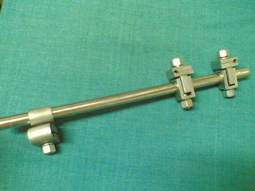
What changed. Parkhill (1894) and Lambotte (1902) independently placed pins in bone and joined them to a frame outside the limb. Lambotte introduced threaded pins but still needed an open reduction. In 1938 Raoul Hoffmann (Geneva) reduced the fracture closed and placed the pins percutaneously.
What was still missing. Pin-track infection and the burden of the frame, which remain the fixator's weak points today.
7A.3.8 Röntgen's X-rays (1895)
The problem. Reduction, alignment and callus were invisible, so healing was judged by touch and by time.
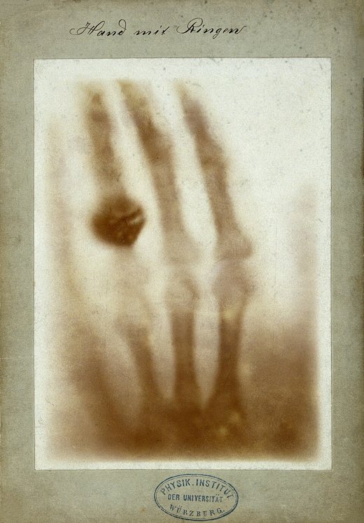
What changed. Wilhelm Röntgen discovered X-rays in November 1895. Radiographs let surgeons see the fracture, check the reduction and follow callus.
What was still missing. A radiograph shows mineral, not strength. Radiographic union is not the same as mechanical union.
7A.3.9 Pins and wires (1907–1909)
The problem. Traction could be applied only through the skin, and cerclage wire could not control the fragments of a long bone.
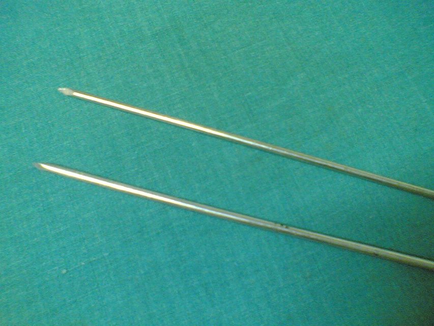
What changed. Steinmann's pin (early 1900s) allowed traction through the bone itself. In 1909 Martin Kirschner introduced the thin wire that still carries his name.
What was still missing. Pins and wires hold small fragments and provide traction, but a long-bone shaft still needed a stronger partner: the plate, and later the nail.
7A.4 Rigid fixation and functional treatment (1925 to the 1960s)
Between the wars, two ideas competed: hold the fracture rigidly, or treat it closed and let the limb work. Both reached maturity in the 1950s and 1960s, and both left biological problems behind.
7A.4.1 Böhler's system of trauma care, and closed treatment (1925–1950)
The problem. Fracture care had no organized system, and long immobilization left the uninjured limb and joints idle.
What changed. Lorenz Böhler opened Vienna's accident hospital in December 1925. His principles were rapid diagnosis, painless reduction, immobilization of the injured part, and active movement of everything else. John Charnley's 1950 textbook on the closed treatment of common fractures carried the same philosophy forward.
What was still missing. Closed treatment could not reliably control unstable fractures, such as those of the femoral shaft.
7A.4.2 Küntscher's intramedullary nail (Kiel) (1939)
The problem. Even with traction, femoral shaft fractures meant weeks of inactivity, and the plates of the time were weak and needed wide exposure.
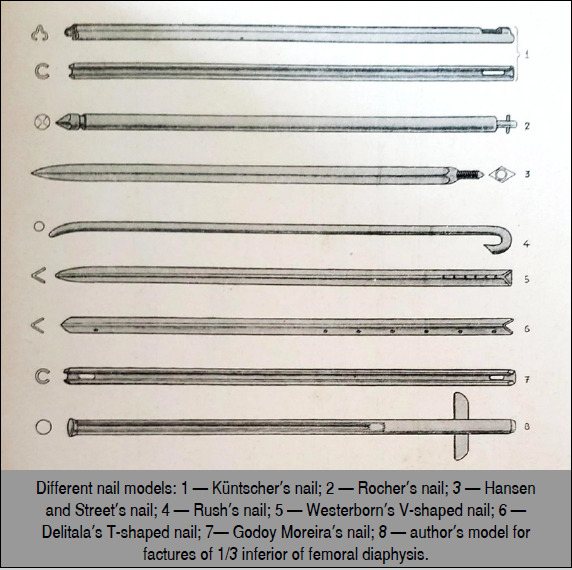
What changed. Intramedullary devices had a long prehistory: Aztec surgeons reportedly used them in the 16th century, and ivory pegs appeared in the late 19th. Gerhard Küntscher succeeded in November 1939 with a rod driven down the medullary canal, without opening the fracture. He reported 39 cases in 1939 and 107 war cases in 1947. Early nails were V-shaped or triangular, later clover-leaf for better rotational stability. Because the nail shares load with the bone, soldiers could return to activity within weeks. He worked closely with the engineer Ernst Pohl, and the Second World War spread the technique around the world despite harsh early criticism.
What was still missing. The unlocked nail worked well in simple fractures of the narrow isthmus of the femur or tibia. In wide-canal or comminuted fractures it gave shortening and bad union. Surgeons tried larger, reamed nails and, above all, ways of locking the nail.
7A.4.3 Flexible pins: Rush and Ender (1930s–1970s)
The problem. A rigid nail needs a canal that fits it, and usually reaming. Smaller bones and fragile older patients needed something gentler.
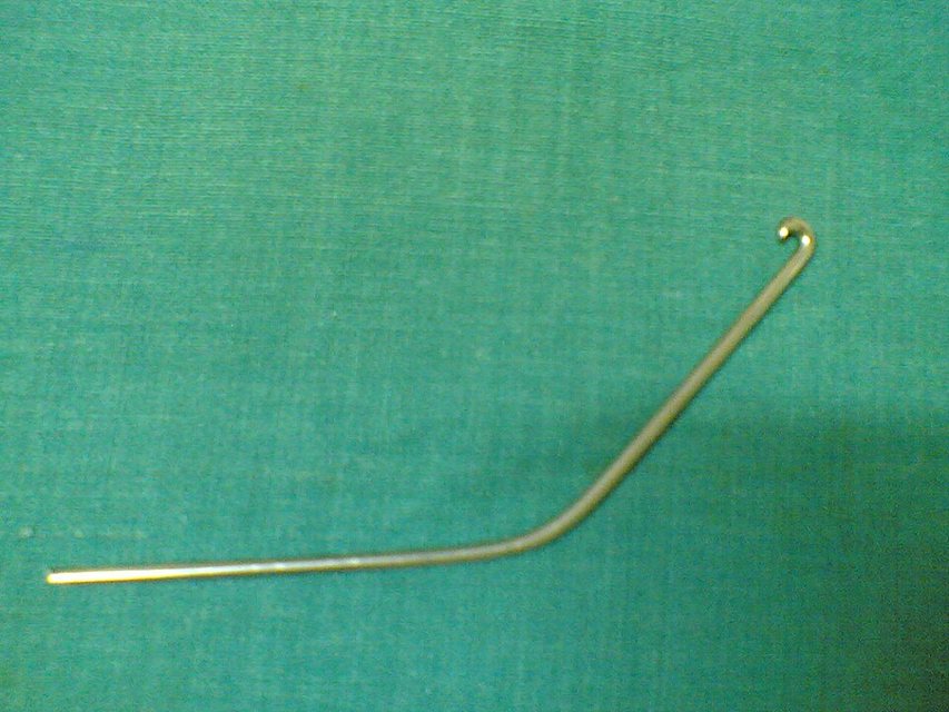
What changed. The Rush brothers developed a slender curved pin driven longitudinally into the canal, which springs against the canal walls to hold the fragments. In Vienna in 1970, Ender applied bundles of elastic pins, inserted from the femoral condyle, to trochanteric fractures in older patients.
What was still missing. Elastic pins do not lock, so rotation and shortening are poorly controlled in unstable fractures. In the 1980s Ender pins were compared with the compression hip screw for unstable intertrochanteric fractures, and the screw, and later the nail, became the mainstay.
7A.4.4 The hip: from screws to sliding screws and cephalomedullary nails (1931–1980s)
The problem. Hip fractures were mostly treated without surgery. In 1883 Senn argued that every femoral neck fracture should be operated on, but few agreed.
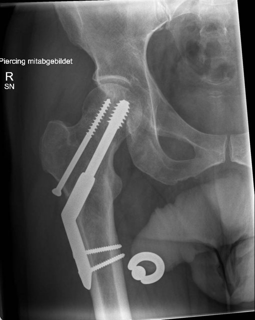
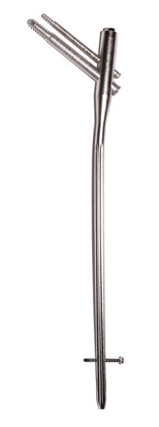
What changed. Langenbeck used a silver screw for a femoral neck fracture in 1858, and König repeated it under antisepsis in 1875. In 1931 Smith-Petersen reported open reduction and fixation with a trifin nail, and guide-wire aids (Johansson 1932, Wescott 1934) made placement practical. The American Academy of Orthopaedic Surgeons endorsed the trifin nail in 1941. Trochanteric fractures were treated with a sliding screw in a side plate, and later with a nail carrying a screw into the femoral head (a cephalomedullary nail).
What was still missing. In 1976 the British Medical Research Council found the trifin nail unsuitable for displaced neck fractures, and cannulated screws (Asnis, 1980) replaced it. Hip nails still fail for several reasons, and their designs are still being refined.
7A.4.5 Bone banks (1940s)
The problem. Autograft is limited in quantity and costs the patient a second operative site.
What changed. Allograft use had waned, but bone banking became a reality during and after the Second World War. The National Naval Tissue Bank was established in Bethesda and small banks opened in hospitals worldwide. Fragments of cortical or cancellous bone augmented spinal fusions, filled cysts and scaffolded delayed union and nonunion.
What was still missing. Banked bone supplies a scaffold, not living cells. Screening, processing and traceability became the next problem.
7A.4.6 Compression: Danis and the "coapteur" (1948–1949)
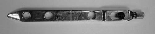
The problem. Bone ends resorb, so a rigid plate without compression left a gap and delayed healing. Slotted plates (Eggers, 1948) let screws slide to follow the collapse but were too weak.
What changed. Robert Danis (Brussels) compressed the fragments with a plate he called the coapteur. Motion was abolished and bone healed without callus. He named this "soudure autogène" (autogenous welding); today we call it primary bone healing.
What was still missing. The idea needed a reproducible system: standard instruments, training and evidence.
7A.4.7 The AO: compression becomes a system (1958–1970s)
The problem. Implants were improvised, results depended on individual skill, and fractures were still immobilized for long periods.
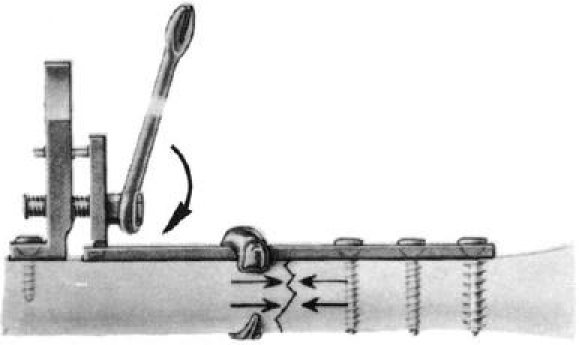
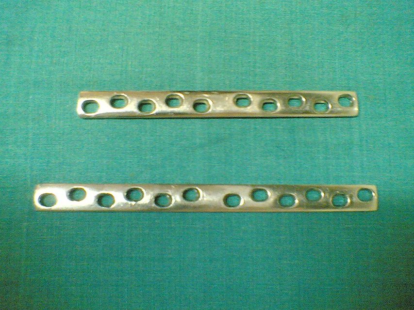
What changed. Maurice Müller met Danis in 1950 and helped found the AO in Switzerland in 1958. The first Davos course (December 1960) taught standardized instruments and four principles: anatomic reduction, stable fixation, preserved blood supply and early active mobilization. A tensioner plate (1965) gave way to the dynamic compression plate, whose oval holes resemble Bagby and Janes's 1958 design. Malunion became uncommon and neighboring joints could move at once.
What was still missing. Rigidity was bought with exposure. Cortical porosis under the plate, delayed bridging and refracture after plate removal followed. Callus, the usual sign of healing, was absent, and its appearance was read as instability.
7A.4.8 Urist and bone induction (1965)
The problem. The fate of grafts had been argued for two centuries, and no one knew what signal made a graft form bone.
What changed. In 1965 Marshall Urist showed that demineralized bone matrix implanted in muscle induces new bone. He named the active factors bone morphogenetic proteins in 1971.
What was still missing. Purifying the proteins and delivering them took decades. Finding a carrier proved harder than expected, and a signal without a carrier, a viable bed and stable fixation does not form reliable bone.
7A.4.9 Functional bracing (1967)
The problem. A full cast immobilizes the joints above and below a fracture and keeps the patient off the limb.
What changed. Sarmiento's functional below-the-knee cast for tibial fractures (1967) allowed early weight bearing and left the knee free.
What was still missing. It suits stable patterns and needs careful watching of alignment. It also depends on callus, so it belongs to the indirect healing route.
7A.5 Biology, regeneration and reconstruction (1968 to the present)
By the 1970s the mechanical problems were largely solved. The failures that remained were biological: devitalized bone, nonunion, bone defects and infection. Progress therefore turned to blood supply, regeneration and reconstruction.
7A.5.1 Locked nails and reaming (1968–1990s)
The problem. Unlocked nails suited only simple isthmic fractures, and in the 1960s plating pushed nailing aside.
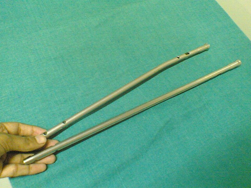
What changed. In 1953 Modny and Bambara described a multiperforated nail with crossed screws for axial and rotational control. In 1968 Küntscher presented the "detensor" nail, soon called the interlocking nail, with one crossing screw at each end. By the 1970s the modern interlocking design had spread worldwide. Flexible reaming, image intensification and titanium nails followed. Locked nailing showed that flexible fixation without precise reduction heals reliably, a direct challenge to the doctrine of rigidity. A footnote: a São Paulo thesis of 1952 had already described a distal locking device, but it was never indexed in a journal.
What was still missing. Reaming and strong implants disturbed the blood supply, and infection rose, especially in open fractures. Unreamed nailing was promoted as "biological osteosynthesis".
7A.5.2 Vascularized bone transfer: the free fibula (1975)
The problem. Large segmental defects, and irradiated or infected beds, could not be reconstructed with conventional grafts, which depend on a viable recipient bed.
What changed. In 1975 Taylor described the free fibula flap. Since then many bone-containing flaps have been described, each chosen for its vascular pedicle and the defect.
What was still missing. The transferred bone must still unite with the host at its junctions, and the microsurgical demand and donor-site cost are considerable.
7A.5.3 Ilizarov: regeneration by tension (1951 → 1980s)
The problem. Infected nonunions, bone defects and deformities defeated plates and nails, or could not be treated with them.
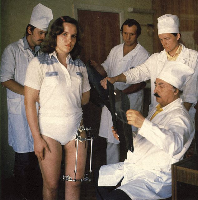
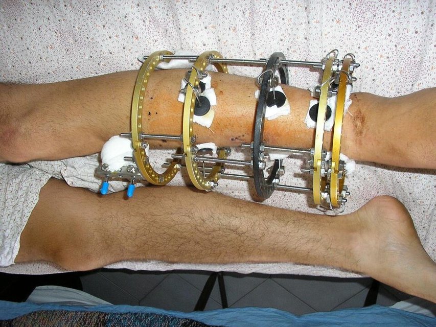
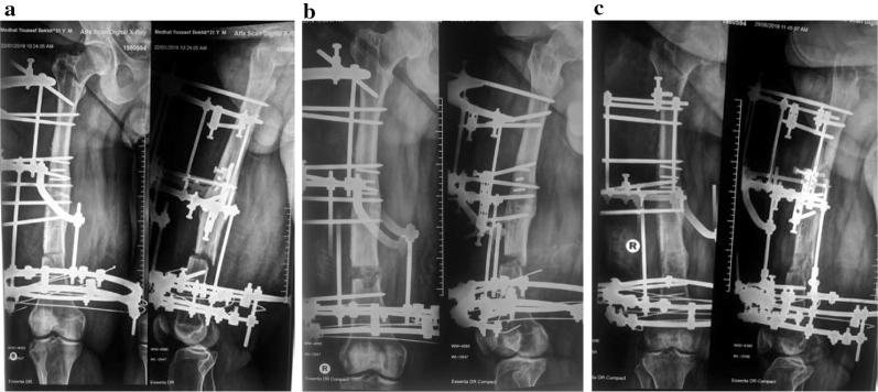
What changed. Gavriil Ilizarov (Kurgan, Siberia) began in 1951 with a circular frame of tensioned thin wires, which allowed controlled motion and early loading. Reportedly after a patient lengthened the frame instead of shortening it, he found that slow tension regenerates bone: distraction osteogenesis. The Olympic high jumper Valeriy Brumel (1968; more than twenty failed operations, then 3.5 cm of new bone) and the explorer Carlo Mauri (1980) were among his patients. The method reached Western countries in the early 1980s and the United States in 1987.
What was still missing. Months in a frame, pin and wire site infection, and a heavy burden on the patient.
7A.5.4 Biological fixation and the internal fixator (1990s–2000s)
The problem. Anatomic reduction of every fragment, whether with a rigid plate or by reaming, cost blood supply. Delayed union and infection followed.
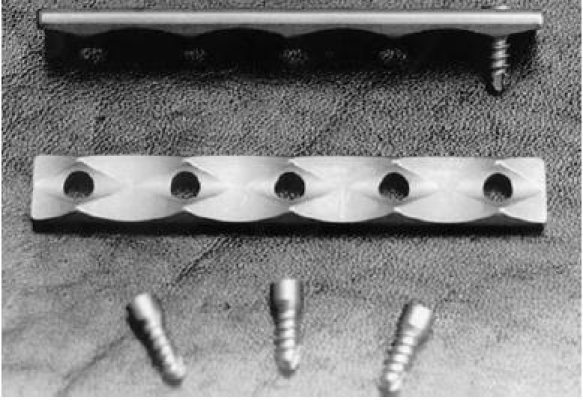
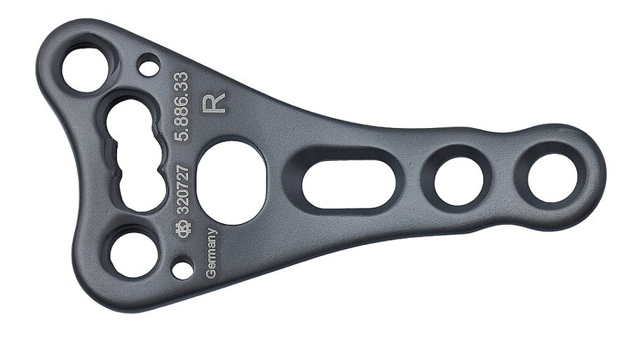
What changed. Surgeons bridged the fracture zone, using indirect reduction and limited exposure. Then came the internal fixator: locking screws thread into the plate (point-contact fixator, LISS, LCP), so the plate splints rather than compresses. It need not touch or match the bone and can be inserted percutaneously (MIPO). The flexible construct produces callus.
What was still missing. Excess rigidity can still suppress callus, so "dynamic" plating with more controlled motion is the newest idea. The older question is still argued: do limited-contact plates protect cortical perfusion, or is bone loss simply stress shielding?
7A.5.5 Recombinant BMP and cell-based adjuncts (2002 →)
The problem. Autograft remained the reference standard, but its supply was limited and its harvest costly, and BMP research had shown that a bone-forming signal could be supplied.
What changed. Recombinant BMP-2 on an absorbable collagen sponge (INFUSE) was approved by the FDA on 2 July 2002 for one-level anterior lumbar interbody fusion, and on 13 February 2026 for transforaminal lumbar interbody fusion. Over the last four decades, marrow aspiration was also developed to treat nonunion, for example in Hernigou's 2005 series of 60 tibial nonunions.
What was still missing. Approval is specific to a product, a device combination and an indication, and the evidence for most adjuncts is variable.
7A.5.6 Where nails meet frames (2000s–now)
The problem. Frames give reliable lengthening and deformity correction, but they are burdensome and pin sites become infected.
What changed. Lengthening over a nail, or lengthening then nailing, lets the frame come off early. Self-distracting, magnetically driven intramedullary nails now lengthen bone without external hardware.
What was still missing. Complications remain, and Ilizarov's principles are still the cornerstones of every lengthening method.
7A.6 The facial skeleton (1886 to the present)
Facial fracture fixation followed the same arc as the limb, with its own constraints: thin bone, teeth, the occlusion, and scars that everyone can see.
7A.6.1 Plates for the mandible: from Hansmann to Luhr (1886–1968)
The problem. Jaw fractures were held with wires, arch bars and weeks of maxillomandibular fixation. Plating had a high complication rate and was not generally accepted, and limb implants did not suit facial anatomy.
What changed. After Danis's compression principle reached the AO, Luhr (Göttingen) designed a self-compressing plate for the mandible and performed the first compression plating in the maxillofacial region around 1967–68.
What was still missing. Corrosion-prone steel, and plates of the wrong dimensions for a thin facial skeleton.
7A.6.2 Miniplates, reconstruction plates and titanium (1970s onward)
The problem. The jaws had to be wired shut for weeks.
What changed. Michelet and Champy introduced small monocortical miniplates without compression for mandibular fractures, and later for the midface. Reconstruction plates bridged defects and fixed grafts. Vitallium, then titanium, replaced corrosion-prone steel. Postoperative maxillomandibular fixation could be avoided, which also transformed orthognathic surgery.
What was still missing. Each plate must match the load it carries: a miniplate shares load across a reduced fracture, while a reconstruction plate must bear it.
7A.6.3 Bone from the periosteum: cleft periosteoplasty (1700s–1960s)
The problem. A maxillary cleft needs bone continuity across the alveolus.
What changed. Experiments by Troja, Larghi, Ollier and Langenbeck established the osteogenic power of the periosteum, and the plastic surgeon Tord Skoog turned it into periosteoplasty for maxillary clefts. The technique rests directly on the 250-year-old Duhamel–Ollier question.
What was still missing. Periosteoplasty is one of several options for maxillary clefts, and larger defects still need grafts or vascularized bone.
7A.6.4 Virtual planning and 3D printing (2000s–now)
The problem. Panfacial fractures leave no reliable bony or soft-tissue landmarks, so poor reduction produces a permanent deformity.
What changed. Two- and three-dimensional CT, virtual surgical planning and 3D-printed skull models let the surgeon plan reduction and fixation before the operation.
What was still missing. The plan does not replace judgment. Fractures must still be exposed, reduced and checked against the occlusion, and primary repair is best within about two weeks.
7A.7 Timeline at a glance
| When | Step |
|---|---|
| c. 1600 BCE | Splints and bandages in the ancient world |
| AD 30–1500s | Stiffened bandages |
| 1700s–1867 | Where does new bone come from? |
| 1851 | Plaster of Paris |
| 1860s–1880s | Anesthesia and antisepsis: opening a fracture becomes survivable |
| 1875–1918 | Traction and the Thomas splint |
| 1878–1881 | The first bone allograft |
| 1886–1912 | The first plates and screws |
| 1892 | Wolff: bone adapts to load |
| 1894–1938 | External fixation: keeping hardware out of the wound |
| 1895 | Röntgen's X-rays |
| 1907–1909 | Pins and wires |
| 1925–1950 | Böhler's system of trauma care, and closed treatment |
| 1939 | Küntscher's intramedullary nail (Kiel) |
| 1930s–1970s | Flexible pins: Rush and Ender |
| 1931–1980s | The hip: from screws to sliding screws and cephalomedullary nails |
| 1940s | Bone banks |
| 1948–1949 | Compression: Danis and the "coapteur" |
| 1958–1970s | The AO: compression becomes a system |
| 1965 | Urist and bone induction |
| 1967 | Functional bracing |
| 1968–1990s | Locked nails and reaming |
| 1975 | Vascularized bone transfer: the free fibula |
| 1951 → 1980s | Ilizarov: regeneration by tension |
| 1990s–2000s | Biological fixation and the internal fixator |
| 2002 → | Recombinant BMP and cell-based adjuncts |
| 2000s–now | Where nails meet frames |
| 1886–1968 | Plates for the mandible: from Hansmann to Luhr |
| 1970s onward | Miniplates, reconstruction plates and titanium |
| 1700s–1960s | Bone from the periosteum: cleft periosteoplasty |
| 2000s–now | Virtual planning and 3D printing |
7A.8 Summary
- For most of history a fracture was held still and left to heal. Casts, anesthesia, antisepsis and X-rays made the first operations on bone possible in the late 1800s.
- Early plates failed from corrosion and weakness. Danis showed that a compressed fracture heals without callus, and the AO turned compression into a teachable system.
- Rigid fixation solved the mechanical problem but cost blood supply. Locked nails, bridging plates and locking plates moved fixation toward biology.
- The remaining failures were biological: dead bone, defects and infection. The answers were Urist's bone induction, Ilizarov's distraction and Taylor's free fibula.
- The face followed the same path, from wires and weeks of jaw fixation to Luhr's compression plates, Champy's miniplates and virtual planning.
- The 250-year-old question of whether a graft lives or only guides new bone (Duhamel and Ollier) still underlies how grafts are classified today.
References
- Uhthoff HK, Poitras P, Backman DS. Internal plate fixation of fractures: short history and recent developments. J Orthop Sci 2006;11:118–126.
- Perren SM. Evolution of the internal fixation of long bone fractures. The scientific basis of biological internal fixation: choosing a new balance between stability and biology. J Bone Joint Surg Br 2002;84:1093–1110.
- Broos PL, Sermon A. From unstable internal fixation to biological osteosynthesis. A historical overview of operative fracture treatment. Acta Chir Belg 2004;104:396–400.
- Augat P, von Rüden C. Evolution of fracture treatment with bone plates. Injury 2018;49 Suppl 1:S2–S7.
- Andrianne Y, Hinsenkamp M. Historical review of the treatment of fractures. Contribution of the Belgian surgery to the origin and development of osteosynthesis. Rev Med Brux 2011;32:S30–S37.
- Nakayama DK. The Welsh clan that transformed orthopedic surgery. Am Surg 2019;85:441–448.
- Bekos A, Sioutis S, Kostroglou A, et al. The history of intramedullary nailing. Int Orthop 2021;45:1355–1361.
- Camargo FP, Gaiarsa GP, Camargo OP, et al. The missing link in the history of the locked intramedullary nail. Acta Ortop Bras 2021;29:228–231.
- Buhl CA. 80 years of intramedullary nailing: new facts and information about a milestone in osteosynthesis. Unfallchirurg 2019;122:127–133.
- Russell TA, Klima ML. History and evolution of hip design to decrease failure. J Orthop Trauma 2023;37:S49–S54.
- Mei J. A brief history of internal fixation of femoral neck fracture. Zhonghua Yi Shi Za Zhi 2014;44:101–105.
- Sarmiento A. A functional below-the-knee cast for tibial fractures. J Bone Joint Surg Am 1967;49:855–875.
- Hosny GA. Limb lengthening history, evolution, complications and current concepts. J Orthop Traumatol 2020;21:3.
- Gubin AV, Borzunov DY, Malkova TA. The Ilizarov paradigm: thirty years with the Ilizarov method, current concerns and future research. Int Orthop 2013;37:1533–1539.
- Hernigou P. Bone transplantation and tissue engineering. Part II: bone graft and osteogenesis in the seventeenth, eighteenth and nineteenth centuries (Duhamel, Haller, Ollier and MacEwen). Int Orthop 2015;39:193–204.
- Hernigou P. Bone transplantation and tissue engineering, part III: allografts, bone grafting and bone banking in the twentieth century. Int Orthop 2015;39:577–587.
- Hernigou P. Bone transplantation and tissue engineering, part IV. Mesenchymal stem cells: history in orthopedic surgery. Int Orthop 2015;39:807–817.
- Di Matteo B, Tarabella V, Filardo G, et al. An orthopaedic conquest: the first inter-human tissue transplantation. Knee Surg Sports Traumatol Arthrosc 2014;22:2585–2590.
- Meikle MC. On the transplantation, regeneration and induction of bone: the path to bone morphogenetic proteins and other skeletal growth factors. Surgeon 2007;5:232–243.
- Grgurevic L, Pecina M, Vukicevic S. Marshall R. Urist and the discovery of bone morphogenetic proteins. Int Orthop 2017;41:1065–1069.
- Sparks DS, Saleh DB, Rozen WM, et al. Vascularised bone transfer: history, blood supply and contemporary problems. J Plast Reconstr Aesthet Surg 2017;70:1–11.
- Lazzeri D, Gatti GL, Romeo G, et al. Bone regeneration and periosteoplasty: a 250-year-long history. Cleft Palate Craniofac J 2009;46:621–628.
- Luhr HG. The development of modern osteosynthesis. Mund Kiefer Gesichtschir 2000;4 Suppl 1:S84–S90.
- US Food and Drug Administration. INFUSE Bone Graft: Summary of Safety and Effectiveness Data, P000058/S096.
Early dates differ between sources, so approximate periods are given.