2.1 Introduction
Under the microscope, bone tissue is not a uniform solid. It is built in levels, one inside the other. The strength of a bone depends on every level, not only on how much mineral it contains (Rho 1998; Seeman and Delmas 2006).
Flowchart: Levels of organization of bone, from large to small
- Whole boneshape and size
- Cortical and cancellous bonedense shell, inner network
- Osteons and trabeculaethe structural units
- Lamellaelayers about 3 to 7 µm thick
- Collagen fibrils and mineral crystalsthe material
Bone tissue is described in two separate ways:
Flowchart: Two ways of describing bone tissue
- Bone tissue
- By architecture
- Cortical (compact)dense; made of osteons
- Cancellous (trabecular)a network of struts in marrow
- By matrix organization
- Wovenimmature; collagen at random
- Lamellarmature; collagen in ordered layers
- By architecture
These are not four types of bone. They are two separate descriptions. A mature trabecula is cancellous and lamellar. A fracture callus is woven and later becomes lamellar.
2.2 Cortical and cancellous bone
Every bone has a shell of cortical bone. Inside most bones there is cancellous bone, with marrow in the spaces.
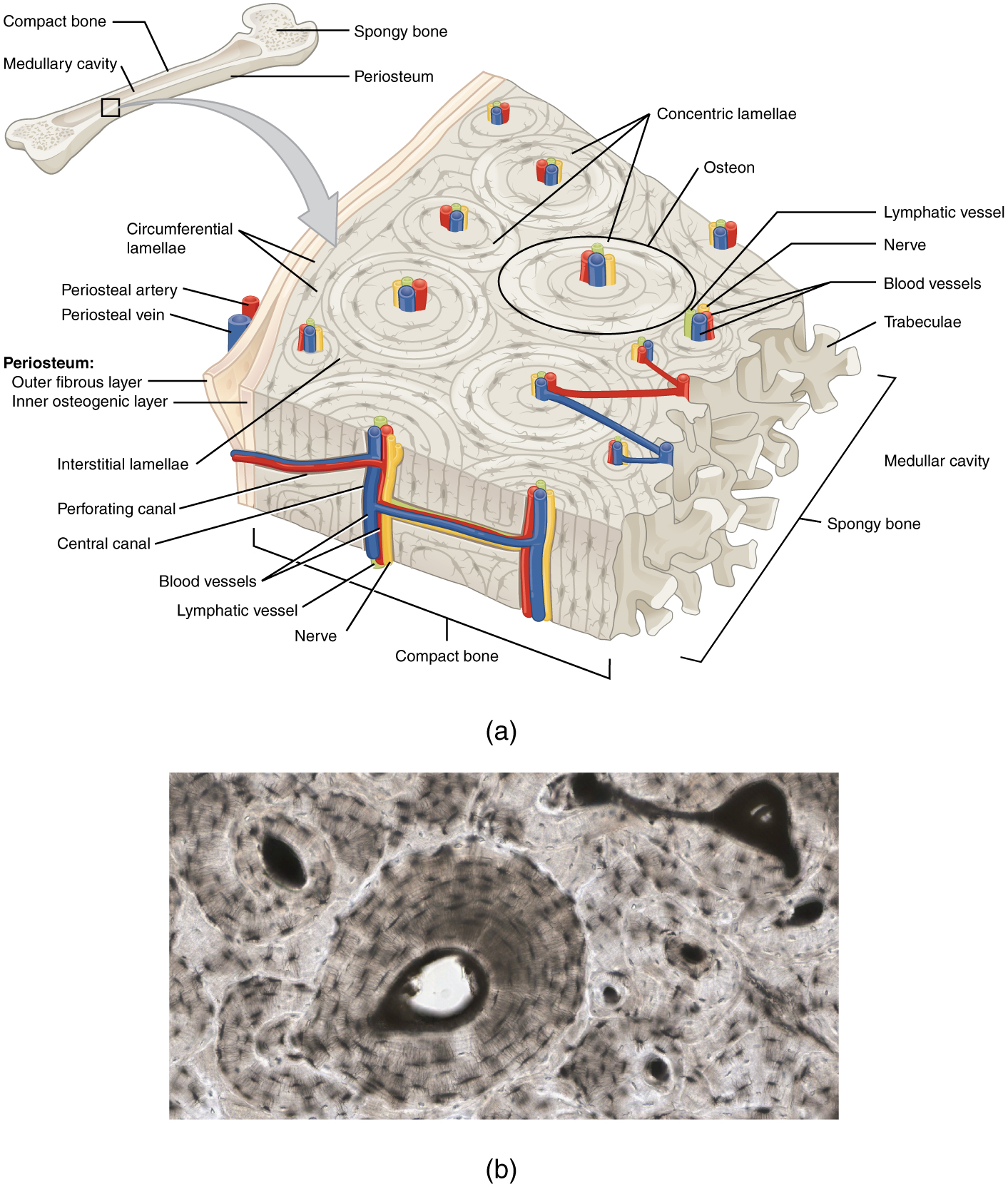
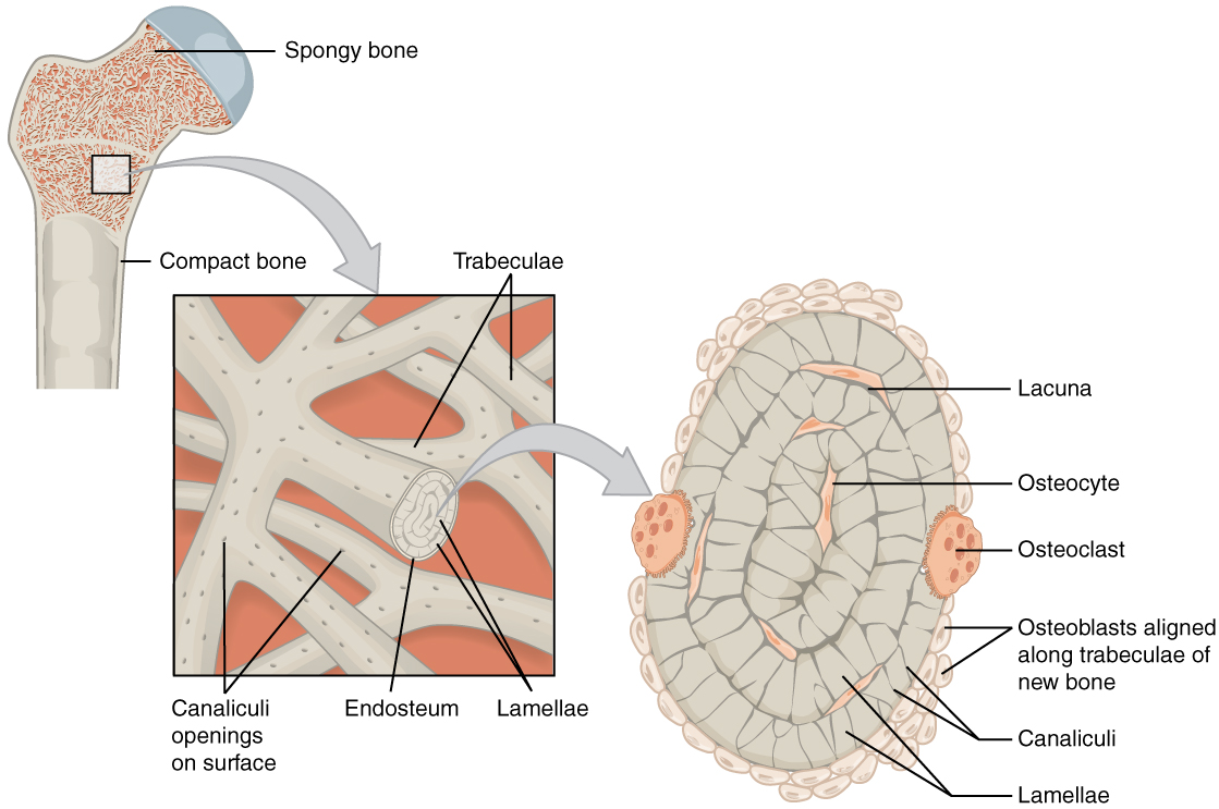
| Cortical bone | Cancellous bone | |
|---|---|---|
| Share of skeletal mass | About 80% | About 20% |
| Structure | Osteons packed together | Plates and rods (trabeculae) separated by marrow |
| Porosity | Low, about 5 to 10% | High, about 50 to 90% |
| Surface area for each unit of volume | Small | Large |
| Turnover | Slow, about 3% a year | Fast, about 25% a year |
| Where it is thickest | Shafts of long bones, the mandible, the tables of the skull | Metaphyses, vertebral bodies, iliac crest, the diploë |
| Main mechanical role | Resists bending and twisting | Spreads load near joints; supports the cortex from inside |
Both types have a mechanical role and both are metabolically active. Cancellous bone turns over faster because it has more surface for cells to work on.
2.3 Cortical bone: lamellae and osteons
In mature cortical bone the matrix is laid down in thin layers called lamellae. They are arranged in four ways:
- Outer circumferential lamellae, just under the periosteum, running around the whole bone.
- Inner circumferential lamellae, lining the marrow cavity.
- Osteonal (concentric) lamellae, forming the osteons.
- Interstitial lamellae, the fragments between osteons. They are the remains of older osteons that were partly removed.
2.3.1 The osteon
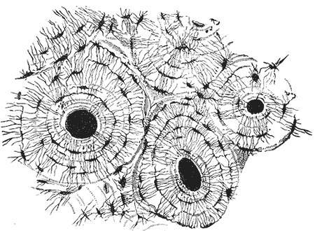
The osteon (Haversian system) is the structural unit of cortical bone. It is a cylinder, about 0.2 mm wide, running mainly along the long axis of the bone. It consists of:
- A central (Haversian) canal containing one or two small vessels and a nerve.
- Concentric lamellae around the canal, usually 4 to 20 of them.
- Lacunae: small spaces between the lamellae, each containing one osteocyte.
- Canaliculi: very fine channels that join the lacunae to each other and to the central canal.
- A cement line at the outer edge. It marks where the osteoclasts stopped when they cut the tunnel in which the osteon was built.
Volkmann (perforating) canals run across the bone, at right angles to the osteons. They connect the central canals with each other, with the periosteum and with the marrow. Unlike central canals, they are not surrounded by concentric lamellae.
2.3.2 Primary and secondary osteons
| Primary osteon | Secondary osteon | |
|---|---|---|
| How it forms | Bone is laid down around an existing vessel | Osteoclasts first cut a tunnel through existing bone; osteoblasts then fill it |
| When | During growth | Throughout life, during remodeling |
| Cement line | No | Yes |
| Importance | Builds new cortex | Replaces old or damaged cortex; is the mechanism of direct (primary) fracture healing |
A secondary osteon is made by a cutting cone of osteoclasts that bores forward, followed by a closing cone of osteoblasts that fills the tunnel from the outside inward, layer by layer, until only the central canal is left (Parfitt 1994). When a fracture is compressed with a plate and there is no movement, these cutting cones cross the fracture line and join the two ends directly, without callus.
2.4 Cancellous bone: trabeculae
Trabeculae are plates and rods, about 0.1 to 0.2 mm thick. Their quality depends on:
- Thickness and number
- Spacing between them
- Connectivity: how well they join each other
- Orientation: they line up along the lines of load
Trabeculae are thin enough that their cells get nutrients from the marrow on their surfaces. They do not need central canals.
Two bones can contain the same amount of mineral and still differ in strength. If trabeculae are perforated and lose their connections, a load path is lost and the remaining struts carry more force. This is what happens in osteoporosis, and it is why bone density alone does not predict fracture (Seeman and Delmas 2006).
2.5 Woven and lamellar bone
| Woven bone | Lamellar bone | |
|---|---|---|
| Speed of formation | Fast | Slow |
| Collagen | Interlacing fibers at random | Parallel fibers in each lamella; the direction changes from one lamella to the next |
| Cells | Many, large, at random | Fewer, evenly spaced |
| Mineral | Patchy | Even |
| Strength | Weak | Strong |
| Where it is found | Fetus, fracture callus, distraction gap, around a bone graft; also in fibrous dysplasia and Paget disease | Almost all of the adult skeleton |
Woven bone is the body's first response whenever bone must be made quickly (Shapiro and Wu 2019). It is a normal stage, not a disease. It does not turn into lamellar bone by rearranging its collagen. It is removed by osteoclasts and replaced by new lamellar bone. This replacement takes months.
This explains a common finding after reconstruction: the X-ray may show that a fracture, an osteotomy or a graft junction is bridged long before the new bone is strong. The bridge is woven bone, and it still has to be remodeled into lamellar bone.
2.6 The osteocyte network
Osteocytes make up more than 90% of the cells in adult bone (Bonewald 2011). Each sits in its lacuna and sends dozens of fine processes through the canaliculi to touch its neighbors and the cells on the bone surface.
- The network carries nutrients and waste through the fluid in the canaliculi.
- When bone is loaded, this fluid moves. The osteocytes sense the movement. This is how bone senses load.
- Osteocytes send signals that start bone removal or bone formation.
This network is not the same as the blood vessels. The vessels run in the central and perforating canals. The canaliculi carry tissue fluid from the vessels to the cells. If the vessels are lost, the osteocytes die within days, even though the canaliculi are still there. Empty lacunae under the microscope are the sign of dead bone.
2.7 Structure and strength
Bone is anisotropic: its strength depends on the direction of the load, because its osteons and collagen are aligned. Cortical bone is strongest in compression along its long axis, weaker in tension, and weakest in shear (Rho 1998).
Strength also depends on:
- Geometry. Bone placed far from the center of a shaft resists bending and twisting much better than the same amount of bone near the center. This is why a long bone is a tube.
- Porosity. More holes in the cortex mean less strength.
- Matrix quality. Collagen gives toughness; mineral gives stiffness.
- Microdamage. Small cracks build up with use and must be removed by remodeling.
2.8 Microarchitecture and bone grafts
The structure of a graft decides how quickly the host can bring vessels and cells into it.
| Graft | Architecture | What follows |
|---|---|---|
| Cancellous | Open spaces, large surface | Vessels grow in within days. Incorporates quickly. Gives almost no structural support. |
| Cortical | Dense osteons, few openings | Vessels can enter only through existing canals and new tunnels cut by osteoclasts. Incorporates slowly, over months to years. Gives early support, but becomes weaker for a time while it is being remodeled. |
| Corticocancellous | Cortical plate with cancellous bone attached | Some support and some rapid ingrowth |
| Vascularized bone flap | Any of the above, with its own vessels | The osteocytes stay alive. It heals like a fracture. It still needs good fixation and contact with the host bone. |
A dense graft on an X-ray shows only that mineral is present. It does not show that the cells are alive, that the graft has been replaced by new bone, or that both ends have united with the host.
2.9 Summary
- Bone is built in levels, from the whole bone down to collagen and mineral. Strength depends on all of them.
- Cortical and cancellous describe architecture. Woven and lamellar describe matrix organization.
- The osteon is the unit of cortical bone. Secondary osteons are made by cutting cones and closing cones, which is also how direct fracture healing works.
- Trabecular connectivity matters as much as the amount of bone.
- Woven bone is made fast and is weak. It is removed and replaced by lamellar bone over months.
- Osteocytes form a network that senses load. Empty lacunae mean dead bone.
- Cancellous graft incorporates fast; cortical graft incorporates slowly but gives support.
References
- Rho JY, Kuhn-Spearing L, Zioupos P. Mechanical properties and the hierarchical structure of bone. Med Eng Phys 1998;20:92–102.
- Seeman E, Delmas PD. Bone quality: the material and structural basis of bone strength and fragility. N Engl J Med 2006;354:2250–2261.
- Parfitt AM. Osteonal and hemi-osteonal remodeling: the spatial and temporal framework for signal traffic in adult human bone. J Cell Biochem 1994;55:273–286.
- Shapiro F, Wu JY. Woven bone overview: structural classification based on its integral role in developmental, repair and pathological bone formation throughout vertebrate groups. Eur Cell Mater 2019;38:137–167.
- Bonewald LF. The amazing osteocyte. J Bone Miner Res 2011;26:229–238.