10B.1 Introduction
This chapter follows one operation from start to finish.
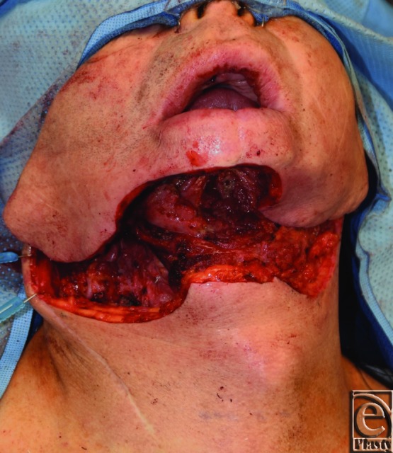
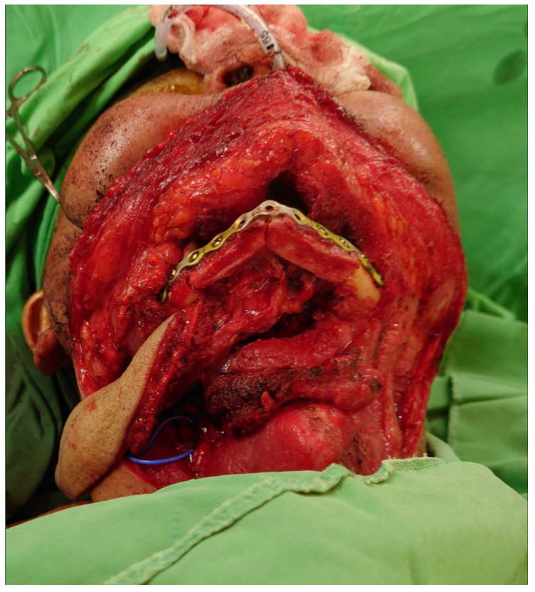
The patient has a tumor of the mandible that also involves the skin of the cheek or chin. The resecting team will remove a full segment of the mandible, the mucosa inside the mouth and the skin outside. This is called a composite through-and-through defect: bone, inner lining and outer skin are all missing.
The plastic surgery team reconstructs the defect at the same operation. We use a free fibula osteocutaneous flap. "Free" means that the flap is detached completely and its vessels are joined to vessels in the neck by microsurgery. "Osteocutaneous" means that the flap contains bone and skin.
The fibula flap for the mandible was described by Hidalgo in 1989. It is now the most used bone flap for the mandible. In a review of 9,499 mandibular reconstructions with bone flaps, 6,178 were fibula flaps (Brown 2017).
Flowchart: Overview of the operation
- 1. Assessment and planningpatient, leg, defect, skin
- 2. Resectionby the resecting team
- 3. Raising the flapat the same time, second team
- 4. Shaping the boneosteotomies and plate
- 5. Inset and fixation
- 6. Vessel anastomosis
- 7. Skin closureface, neck and leg
- 8. Care after surgerymonitoring, complications
10B.2 The defect
10B.2.1 Anatomy of the mandible
The mandible is a U-shaped bone. It is the only moving bone of the face. It is described in regions, from the midline backward:
Flowchart: Regions of the mandible, from the midline to the joint
- Symphysismidline, between the canines
- Bodycarries the premolar and molar teeth
- Anglewhere body meets ramus
- Ramusthe vertical part
- Condyleforms the joint with the skull
- The coronoid process is the front projection at the top of the ramus. The temporalis muscle attaches to it.
- The alveolar part is the upper border of the body, which holds the teeth. It shrinks after the teeth are lost.
- The inferior alveolar nerve and artery enter the inner side of the ramus at the mandibular foramen and run forward in a canal inside the body. The nerve leaves at the mental foramen as the mental nerve, which gives feeling to the lower lip and chin. A segmental resection behind the mental foramen removes this nerve, so the lip is numb on that side.
- The facial artery and vein cross the lower border of the mandible just in front of the masseter muscle. This is where we find them as recipient vessels.
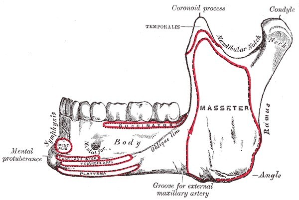
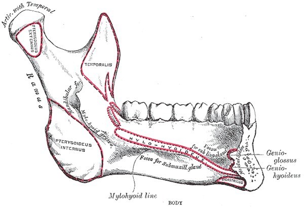
The muscles explain what happens when a segment is removed. Each remaining piece is pulled by the muscles still attached to it.
| Muscles | Attachment | Effect after a segmental resection |
|---|---|---|
| Masseter, medial pterygoid, temporalis (the closing muscles) | Ramus, angle and coronoid process | The posterior piece (ramus and condyle) is pulled upward, forward and inward |
| Lateral pterygoid | Neck of the condyle | The condyle is pulled forward and inward, out of its socket |
| Geniohyoid, genioglossus, digastric, mylohyoid | Inner side of the symphysis and body | The anterior piece is pulled downward and backward. If the symphysis is removed, the tongue loses its front attachment and falls back; this can obstruct the airway. |
This is why the position of the remaining pieces must be recorded and held before the bone is cut, and why the condyles must be checked at the inset.
10B.2.2 What is removed
A segmental mandibulectomy removes the full height of a segment of the mandible, so the mandible is no longer continuous. This is different from a marginal mandibulectomy, where the lower border is kept and continuity is preserved.
In our patient four things are lost. Each must be replaced.
| Tissue lost | What happens if it is not replaced | What replaces it |
|---|---|---|
| Bone | The remaining segments are pulled by the muscles and drift. The bite is lost. The lower face collapses. | Fibula, fixed with a plate |
| Inner lining (mucosa of the floor of mouth, gingiva, cheek) | Saliva leaks into the neck. This causes infection, a fistula, and exposure of the plate and vessels. | Skin paddle of the fibula flap |
| Outer skin (cheek, chin, neck) | The bone, plate and vessels are exposed. | A second skin paddle, or a second flap |
| Soft tissue volume | A hollow contour. Dead space around the bone, which fills with fluid and becomes infected. | Muscle carried with the flap, or a second flap |
Goals of the reconstruction:
- restore the continuity of the mandible;
- keep the condyles in the joint and keep the bite (occlusion) correct;
- restore the height, width and projection of the lower face;
- give a watertight seal between the mouth and the neck;
- cover the outside with stable skin;
- give enough bone for dental implants later.
10B.2.3 Classification of the bone defect
The mandible is described as having four "corners": the two angles and the two canines. The class depends on which corners are removed (Brown 2016).
| Class | Defect | Fibula segments usually needed |
|---|---|---|
| I | Lateral: one angle, not the canine | 1 or 2 |
| II | Hemimandibulectomy: one angle and the canine on the same side | 2 |
| III | Anterior: both canines, neither angle | 2 or 3 |
| IV | Extensive: both canines and at least one angle | 3 or more |
| "c" | Added if the condyle is removed |
The older HCL system is still used: H is a lateral defect that includes the condyle, C is the central segment with both canines, and L is a lateral defect without the condyle (Jewer 1989).
Anterior (central) defects are the most important to reconstruct with bone. Without the anterior arch the chin, lower lip and tongue lose their support. The patient cannot close the mouth, drools, and cannot eat or speak normally. This is called the Andy Gump deformity.
10B.2.4 Why a plate with soft tissue alone is not enough
A simpler option is to bridge the gap with a reconstruction plate and cover it with a soft tissue flap, without bone. The results are poor, especially for anterior defects.
- In 71 patients, a plate without bone failed in 35% of anterior defects and 5% of lateral defects (Boyd 1995).
- In 65 patients with a plate and a soft tissue free flap, the plate became exposed in 46%. Thirty-one percent needed a second operation with a fibula flap (Wei 2003).
The reason is mechanical. A plate without bone carries all the load, at every bite, for life. Metal that is loaded in this way will loosen, break, or cut through the soft tissue. Bone that unites takes the load off the plate.
10B.3 Why the fibula
| Advantages | Disadvantages |
|---|---|
|
|
Results. In 150 consecutive mandible reconstructions with bone free flaps, 90% of them fibula, flap survival was 100% and 97% of the osteotomy sites united (Cordeiro 1999). In the systematic review of 9,499 reconstructions, the failure rate for fibula, radial and scapular flaps together was 3.4% (Brown 2017).
10B.4 Anatomy of the flap
10B.4.1 The bone
- The fibula is on the lateral side of the leg. It carries only a small part of the body weight (commonly quoted as about one sixth). Its main functions are to form the lateral side of the ankle joint and to give attachment to muscles.
- It is 33 to 40 cm long in the adult. It is triangular in cross-section, with thick cortex all around.
- About 6 cm is left at the upper end, to protect the common peroneal nerve and the knee. About 6 to 8 cm is left at the lower end, to keep the ankle stable. The bone between can be taken.
10B.4.2 The compartments of the leg
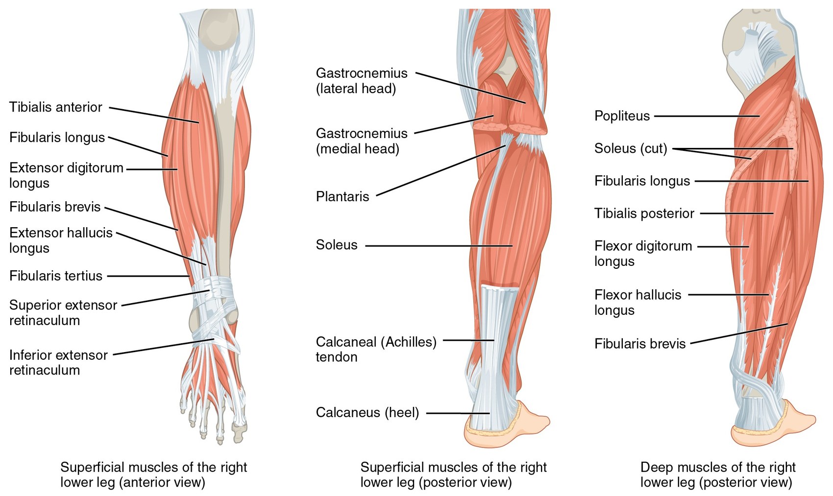
To raise the flap safely, the surgeon must know what lies on each side of the fibula. The leg has four compartments. The fibula touches three of them.
| Side of the fibula | What lies there | What is at risk |
|---|---|---|
| Lateral | Lateral compartment: peroneus longus and peroneus brevis | Superficial peroneal nerve, which runs in this compartment and becomes superficial in the lower third of the leg |
| Anterior | Anterior compartment: extensor digitorum longus, extensor hallucis longus, tibialis anterior | Anterior tibial vessels and deep peroneal nerve, lying on the interosseous membrane |
| Medial | Interosseous membrane, then tibialis posterior | The peroneal vessels themselves |
| Posterior | Deep posterior compartment: flexor hallucis longus (FHL), which arises from the fibula. Behind it, soleus. | Posterior tibial vessels and tibial nerve, which lie more medially |
| Upper end | Neck of the fibula | Common peroneal nerve, which winds around the neck of the fibula. Injury causes foot drop. |
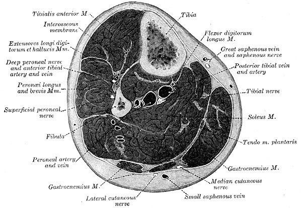
Names. Two sets of names are used for the same structures. Newer texts and some figures use "fibular"; surgeons usually say "peroneal".
| Surgical name | Newer name |
|---|---|
| Peroneal artery | Fibular artery |
| Peroneus longus and brevis | Fibularis longus and brevis |
| Common peroneal nerve | Common fibular nerve |
10B.4.3 The pedicle
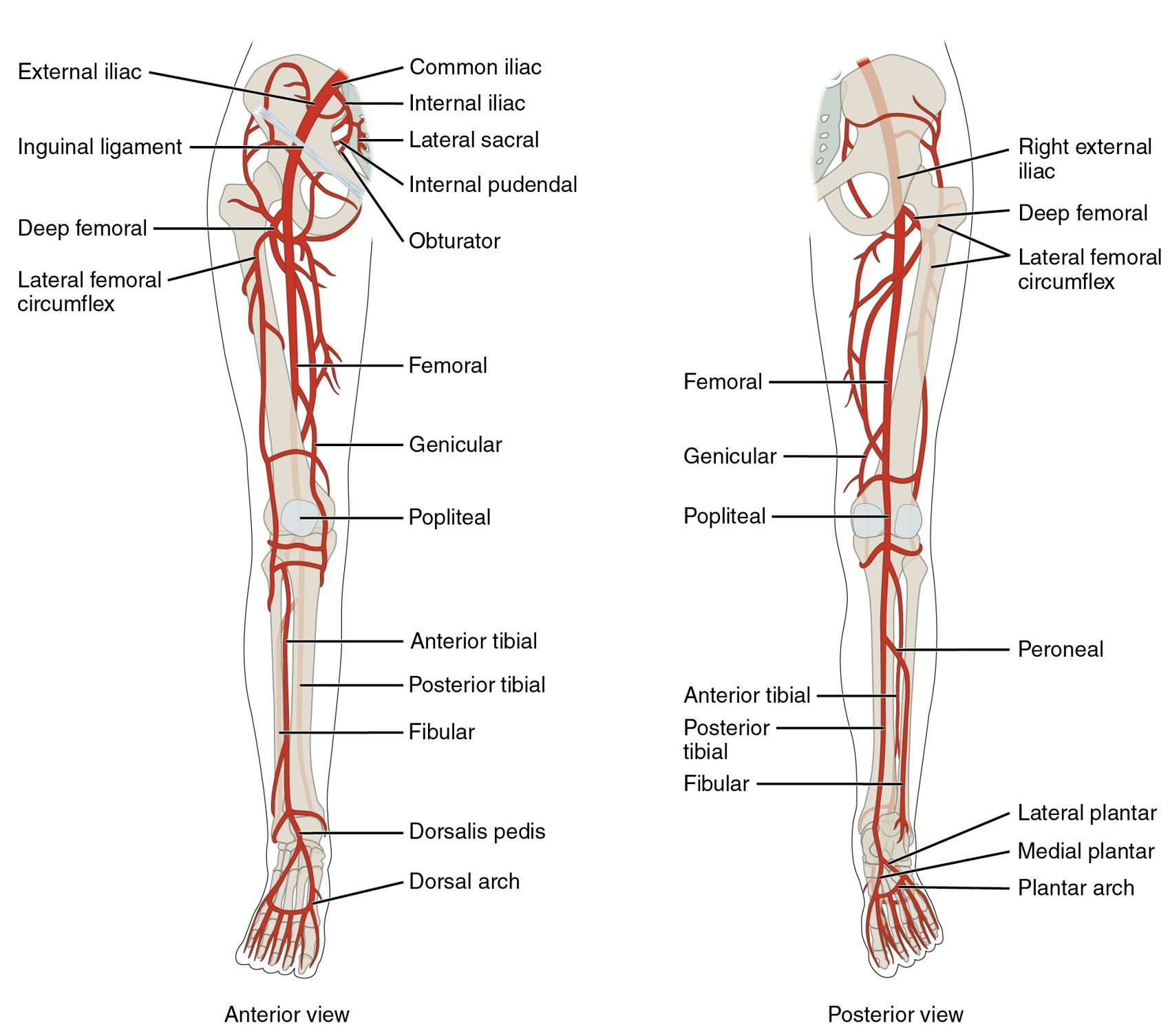
- The pedicle is the peroneal artery with its two veins (venae comitantes).
- The popliteal artery gives off the anterior tibial artery and then continues as the tibioperoneal trunk. The trunk divides into the posterior tibial artery and the peroneal artery. In a cadaver study this division was about 6 cm below the head of the fibula (Gholami 2019).
- The peroneal artery runs down the leg close to the fibula, between tibialis posterior in front and FHL behind.
- The artery is about 1.5 to 3 mm in diameter. The veins are larger.
- The pedicle is short where it begins. It is made longer by using the lower part of the fibula for the reconstruction and removing the unused upper bone from the pedicle. In this way a pedicle of 10 cm or more can be obtained.
10B.4.4 Blood supply of the bone
The peroneal artery supplies the fibula in two ways:
- Endosteal supply: one nutrient artery, which enters the middle third of the bone.
- Periosteal supply: many small branches along the length of the bone, which reach the periosteum directly and through the attached muscle.
When the fibula is cut into segments, the endosteal supply to the segments is interrupted. Each segment then lives on its periosteal supply. This gives two rules for the whole operation: never strip the periosteum, and leave a thin cuff of muscle on the bone.
10B.4.5 Blood supply of the skin
The skin over the lateral leg is supplied by perforators of the peroneal artery. A perforator is a small vessel that leaves the main artery and passes to the skin. There are two types:
- Septocutaneous perforators run in the posterior crural septum, between the peroneal muscles in front and soleus behind. They do not pass through muscle.
- Musculocutaneous perforators pass through soleus or FHL before they reach the skin.
Yu and colleagues mapped 202 perforators in 80 patients, along the line from the head of the fibula to the lateral malleolus (Yu 2011):
| Group | Position on the line | Type | Use |
|---|---|---|---|
| Distal perforators (one to three) | At about one half, two thirds and three quarters of the way down, about 2 cm behind the line | 96% septocutaneous | The main skin paddle is centered here, over the third quarter of the fibula |
| Proximal perforator | At one third of the way down, about 1.5 cm behind the line | 84% musculocutaneous | Can carry a second skin paddle for a through-and-through defect |
In early series the skin paddle was not reliable. Schusterman found that the skin survived in 33% when it was raised on the septum alone, and in 93% when a cuff of soleus and FHL was included (Schusterman 1992). Today the perforators are seen directly before the skin paddle is committed. If a perforator passes through muscle, a cuff of that muscle is taken with it.
The skin can be given sensation by including the lateral sural cutaneous nerve and joining it to a nerve in the mouth (Wei, Chuang and Yim 1994).
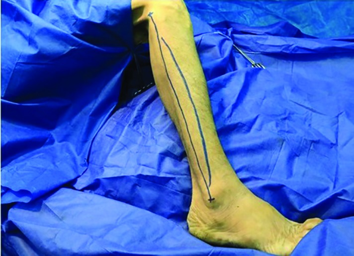
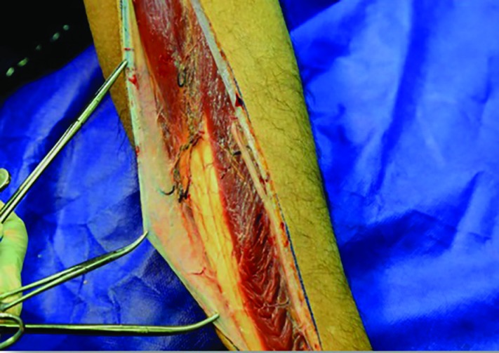
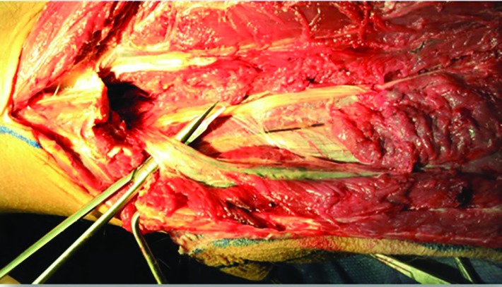
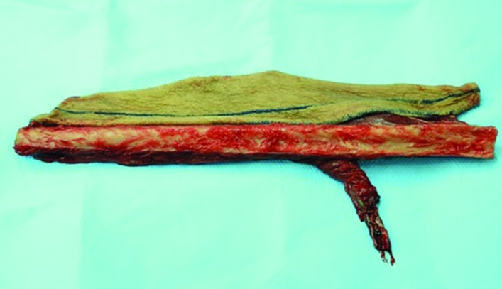
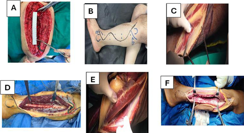
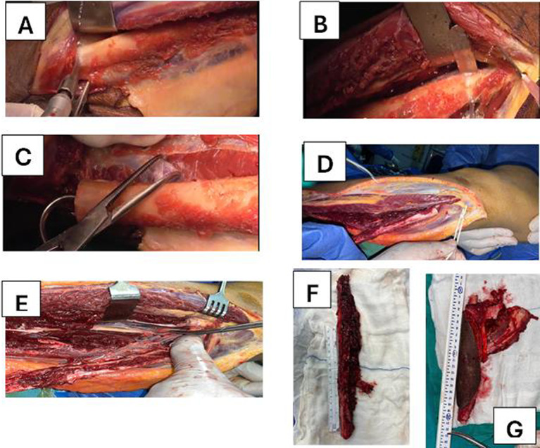
Figures 10B.8 to 10B.11: Gholami et al., World J Plast Surg 2019, CC BY 3.0.
10B.5 Assessment and planning before surgery
10B.5.1 The patient
- General fitness. The operation takes 8 to 12 hours. Assess the heart, lungs, nutrition and diabetes control. Stop smoking.
- Previous treatment. Previous radiotherapy or neck dissection makes the recipient vessels fewer and more fragile, and wound healing poorer.
- Teeth and bite. Record the occlusion. It is the reference for placing the bone.
- Expected treatment after surgery. If radiotherapy is planned, the wounds must heal quickly so that it is not delayed.
10B.5.2 The leg
The purpose is to answer one question: will the foot survive without the peroneal artery?
- History: leg trauma or fracture, previous leg surgery, deep vein thrombosis, claudication, diabetes.
- Examination: palpate the dorsalis pedis and posterior tibial pulses on both sides. Look for varicose veins, edema, ulcers and scars. Check the ankle and great toe movement.
- Imaging: CT angiography or MR angiography of both legs.
What imaging is looking for:
- Peronea arteria magna. The anterior and posterior tibial arteries are small or absent, and the peroneal artery is the main supply to the foot. Taking the fibula flap can then cause ischemia of the foot. It is uncommon but must be excluded.
- Atherosclerosis of the peroneal artery or of the other leg vessels.
- Position of the skin perforators.
Is imaging needed in every patient? In a prospective study of 120 patients with angiography of the donor leg, only 7 of 231 angiograms (3%) showed abnormal vessels. The authors concluded that angiography is needed only when the foot pulses are abnormal or after significant leg trauma (Lutz 1999). Many units now do CT angiography routinely, because it also shows the perforators and gives the images for computer planning. In 40 patients, CT angiography identified 95% of the skin perforators found at operation (Garvey 2012).
Do not use the leg if: the foot depends on the peroneal artery; there is significant peripheral vascular disease; there has been severe trauma or surgery to that leg with damaged vessels; or there is chronic venous disease with poor skin. Choose the other leg or another flap (scapula or iliac crest).
10B.5.3 Planning with the resecting team
The two teams must agree on these points before the operation:
| Question | Why it matters to us |
|---|---|
| How much bone will be removed, and where are the bone cuts? | Decides the length of fibula and the number of segments |
| Will the condyle be kept? | If the condyle goes, the end of the fibula must be shaped and placed in the joint |
| How much mucosa and tongue will be removed? | Decides the size of the inner skin paddle |
| How much skin will be removed? | Decides whether one flap is enough or a second flap is needed |
| What neck dissection is planned, and on which side? | Decides which recipient vessels will be available. Ask the team to preserve the facial artery, the superior thyroid artery and the veins. |
| Will a tracheostomy be done? | The flap and swelling narrow the airway. A tracheostomy is usual. |
10B.5.4 Planning the shape of the bone
The fibula must reproduce the curve of the missing mandible. There are two ways to plan this.
Flowchart: Two ways of planning the bone
- Planning the shape of the fibula
- Conventional method
- The reconstruction plate is bent on the patient's own mandible before the resectionthe screw holes are drilled before the bone is cut
- After the resection the plate is put back in the same holesthe fibula is cut by hand to fit under the plate
- The reconstruction plate is bent on the patient's own mandible before the resectionthe screw holes are drilled before the bone is cut
- Virtual surgical planning
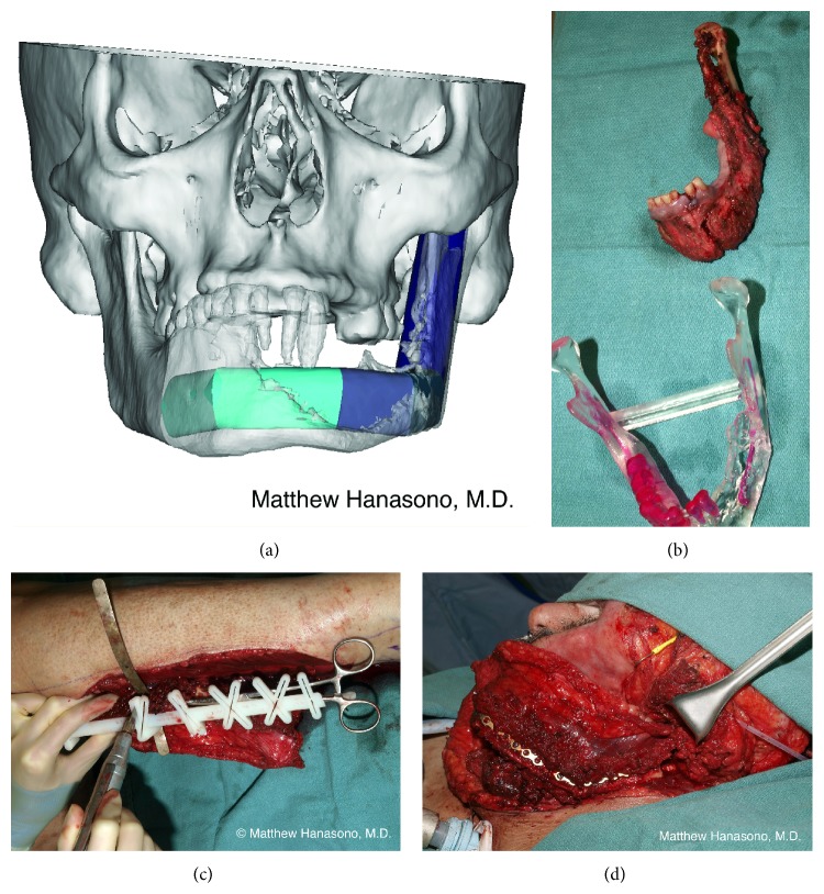
- CT of the jaw and the legthe resection and the fibula segments are planned on a computer
- Printed cutting guides for the mandible and the fibula, and a pre-bent or custom plate
- CT of the jaw and the legthe resection and the fibula segments are planned on a computer
- Conventional method
The conventional method works only when the outer surface of the mandible is normal. If the tumor bulges through the outer cortex, or the mandible is already fractured, the plate cannot be bent on it. Virtual planning is most useful in these cases.
In a comparison of 38 planned cases with matched controls, the mean operating time was 8.8 hours with computer planning and 10.5 hours without. The bony landmarks were also closer to their position before surgery (Hanasono and Skoracki 2013).
Additional note: 3D planning, printed models and cutting guides in current practice
For a planned resection like the one in this chapter, many units now plan the bone work on a computer before the operation and bring printed tools to the operating room. This note describes what is made, how it is used and what the evidence shows.
Terms
| Term | Meaning |
|---|---|
| Virtual surgical planning (VSP) | The resection and the reconstruction are simulated on a three-dimensional computer model made from the patient's CT scans |
| CAD/CAM | Computer-aided design and computer-aided manufacturing: the guides and plates are designed on the computer and then made by a machine |
| 3D printing | Building an object layer by layer from a digital file. Models and guides are printed in resin or nylon (polyamide); plates are printed or milled in titanium. |
| Cutting guide (cutting template) | A printed block that fits the bone in one position only. It has slots that hold the saw at the planned place and angle. |
| Patient-specific plate | A titanium plate made for one patient, already in the planned shape, with planned screw positions |
The workflow
Flowchart: From scan to operation
- 1. Scansthin-slice CT of the face; CT angiography of the legs
- 2. 3D models on the computermandible, tumor, fibula, vessels
- 3. Planning meetingresecting surgeon, plastic surgeon, engineer
- 4. Designguides, plate, models
- 5. Printing and sterilization
- 6. Operation with the guides
- 7. Check after surgeryCT compared with the plan
What is decided at the planning meeting:
- the bone cuts of the resection, with the margin the resecting surgeon needs;
- the number, length and angle of the fibula segments;
- which leg, and how far from the lateral malleolus the first cut is made, so that the skin perforators seen on CT angiography lie over the segments that need skin;
- on which side the pedicle leaves the bone, and which neck vessels it will reach;
- the position of the plate and screws, and of dental implants if they are planned;
- for a defect with skin loss, where the skin paddles must lie in relation to the bone.
What is produced
| Item | What it is for |
|---|---|
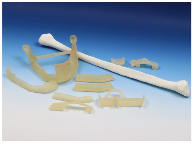
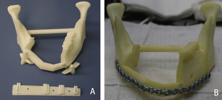
| Printed model of the mandible, as planned after reconstruction | The reconstruction plate is bent on it before surgery. This is possible even when the tumor has destroyed the outer surface of the real mandible. The model is also used to explain the operation to the patient and the team. |
| Mandible resection guides | Fixed to the mandible with screws. They set the two bone cuts. They also carry drill holes, so the screw holes for the plate are made before the bone is cut. The plate then returns the remaining pieces to their original position, and the bite and the condyles are kept. |
| Fibula cutting guide | Fixed to the fibula in the leg, at a planned distance from the ankle. Each slot gives one bone cut at the planned angle. The segments then fit each other without trimming. |
| Positioning template (not always used) | Holds the cut segments in the planned arch while the plate is applied |
| Plate | Either a standard plate bent before surgery on the printed model, or a patient-specific titanium plate |
| Dental implant guide (optional) | Built into the fibula guide, so implants can be placed at the same operation |
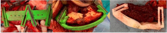
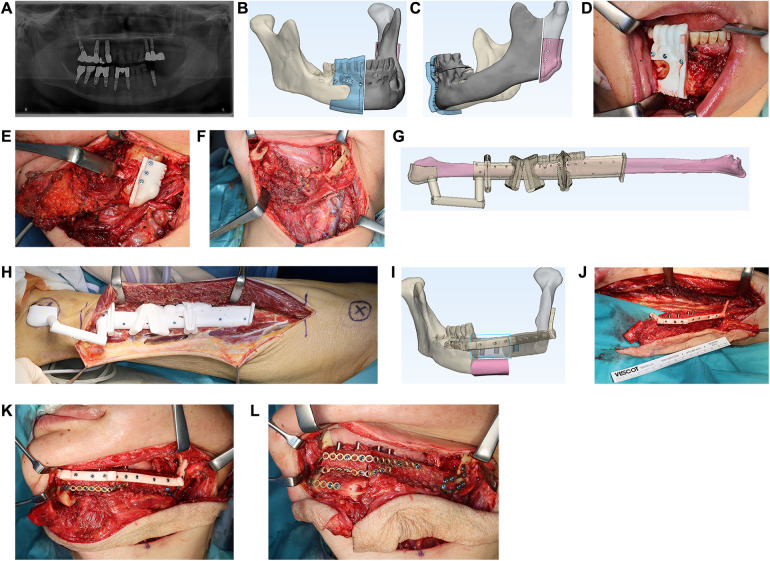
How it changes the operation
- The resection and the flap harvest follow the same plan, so the two teams do not have to wait for each other's measurements.
- The fibula is cut, shaped and fixed to the plate in the leg, while it is still perfused. Only then is the pedicle divided.
- The plate is fixed to holes drilled before the resection. The position of the condyles and the bite does not depend on judgment by eye.
- The steps of marking, raising the flap, joining the vessels and closing the skin are unchanged.
Evidence
| Question | Finding |
|---|---|
| Is the operation shorter? | Yes. In a meta-analysis of 23 studies with 713 patients, planning was associated with shorter operating time and shorter ischemia time (Tang 2019). In one comparison the mean operating time was 8.8 hours with planning and 10.5 hours without (Hanasono and Skoracki 2013). |
| Is the result more accurate? | Yes. Bony landmarks were closer to their position before surgery (mean change 4.1 mm with planning, 6.9 mm without). Segment lengths differed from the plan by a mean of 2.4 mm and angles by 3.5 degrees (Hanasono and Skoracki 2013). |
| Are there fewer complications? | Not shown. In 128 bone flaps, complication rates were not lower with CAD/CAM fixation than with conventional fixation: plate exposure 30% and 19%, incomplete bone union 46% and 33% (Rendenbach 2019). |
| Do patient-specific plates unite as well? | Uncertain. In 89 mandible reconstructions, incomplete union was found in 36% with patient-specific plates and 14% with hand-bent plates (Knitschke 2022). A very rigid plate may take too much load off the bone. |
| Does it delay surgery? | It can. In the same 128 flaps, the time from diagnosis to surgery was 11 days longer with CAD/CAM (34 days and 23 days) (Rendenbach 2019). With planning and printing done inside the hospital, one unit reported no added delay (median 6 days and 8 days) (Lindhardt 2024). |
| What does it cost? | In one unit the extra cost was about US $5,100 per case with a pre-bent plate. After the saved operating time was counted, the net extra cost was about US $1,200 (Zweifel 2015). Costs differ widely between countries and suppliers. |
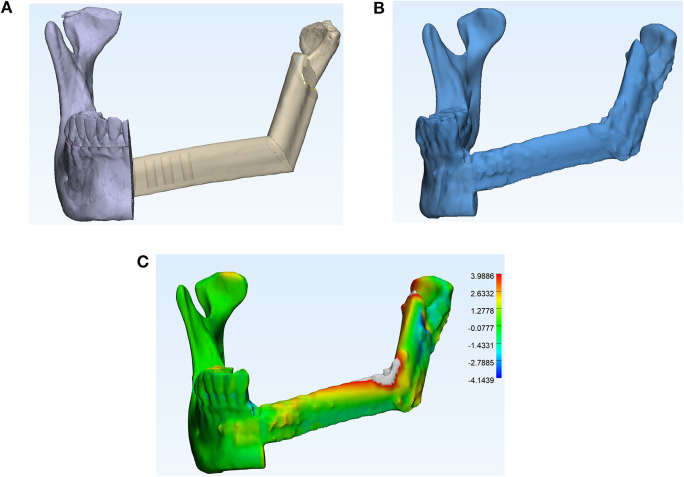
Limits and precautions
- The tumor may grow between the scan and the operation. The planned cuts may then be too close. The resecting surgeon decides the margin on the day. If the cut must move, the guides and a patient-specific plate may no longer fit.
- Always have a conventional plan. Bring a standard reconstruction plate and be ready to shape the fibula by hand. Some units plan a second, wider set of cuts on the same guide.
- The guide must sit exactly. Soft tissue under a guide, or a guide placed a few millimeters off, moves every cut. Clear the bone where the guide sits, but do not strip the periosteum of the fibula more than the guide needs.
- Soft tissue is not planned. The plan fixes the bone. The skin paddle, the perforators and the pedicle still depend on what is found in the leg.
- The scan must be recent and of good quality (thin slices). Metal in the teeth degrades the images.
Who makes it
| Commercial service | In the hospital (point of care) | |
|---|---|---|
| How | The scans are sent to a company. The plan is agreed in an online meeting with its engineer. Guides and plate are delivered. | The hospital has its own software, printer and trained staff |
| Time needed | Usually one to three weeks | Days |
| Cost per case | Higher | Lower per case, after the first investment |
| Titanium patient-specific plates | Available | Usually not; a standard plate is bent on the printed model |
| Responsibility | The company is responsible for the quality of the devices | The hospital is responsible for design, material, sterilization and records |
A simple first step for a unit that is starting. Print only a model of the mandible, corrected on the computer if the tumor has distorted it, for example by mirroring the healthy side. Bend a standard reconstruction plate on the model before surgery and sterilize it. This gives much of the benefit for the shape of the jaw at low cost. Cutting guides can be added later.
10B.5.5 Which leg
The leg decides how the parts of the flap lie in the face. When the lateral surface of the fibula faces outward for the plate, the pedicle lies on the inner (tongue) side. The skin paddle hangs from the posterior border of the bone.
- The side of the leg decides whether the pedicle leaves the bone toward the front or toward the back of the jaw.
- It also decides whether the skin paddle turns easily upward into the mouth or downward onto the neck.
- The choice therefore depends on where the recipient vessels are and whether the skin is needed inside, outside or both (Yagi 2006).
In practice, hold a paper or model template of the flap against the defect before the harvest, and confirm that the pedicle will reach the chosen vessels without a twist. If one leg has abnormal vessels, the other leg is used and the inset is adapted.
10B.6 Planning the skin
In our patient the skin of the face is removed as well as the lining. Two surfaces must be covered. This must be decided before the flap is raised, because it changes how the skin paddle is marked.
In Wei's series of 27 fibula flaps for composite mandibular defects, 18 patients had both a lining and a skin defect. In 5 the fibula skin was enough for both surfaces. In 13 a second free flap or musculocutaneous flap was needed (Wei, Seah and colleagues 1994).
| Option | How it is done | Suitable when | Limitation |
|---|---|---|---|
| 1. One skin paddle, folded | One long paddle. A strip across its middle has the epidermis removed (de-epithelialized). The paddle is folded over the bone: one part inside the mouth, one part outside. | Small lining defect and small skin defect, close together | Folding can kink the perforator. The amount of skin is limited. |
| 2. Two skin paddles on one fibula | Two separate islands of skin, each on its own perforator. They move independently. | Moderate defects of both surfaces. Needs two good perforators. | Depends on the perforator anatomy of that leg |
| 3. Fibula flap and a second free flap | The fibula skin is used for the lining. A second flap, most often the anterolateral thigh flap, covers the outside and adds volume. | Large skin defect, large loss of volume, or extensive resection including tongue or cheek | Longer operation. A second set of recipient vessels is needed, or the second flap is joined to the far end of the peroneal vessels. |
| 4. Fibula flap and a pedicled flap | The fibula skin is used for the lining. A pedicled pectoralis major flap, or a local flap from the neck or chest, covers the outside. | When a second free flap is not possible: no vessels, an unfit patient, or limited operating time | Bulky. Limited reach. Poorer color and contour match. |
Evidence.
- Two paddles. Yang reported 15 double-paddle peroneal flaps used for intraoral and external cover, based on two perforators or two branches of one perforator. This avoided a second free flap (Yang 2000).
- Two free flaps. Wei reported 36 patients with 72 flaps for extensive composite mandibular defects. Complete flap survival was 93%. The mean operating time was 12 hours 10 minutes. In 11 patients the second flap was joined to the far end of the vessels of the first flap (Wei 1999).
- Fibula and pectoralis major. This combination was used in 14 patients with advanced tumors as an alternative to two free flaps (Chen 1999).
Flowchart: Choosing how to cover lining and skin
- Bone, lining and skin are all missing
- Skin defect smalland little loss of volume
- Fibula flap aloneone folded paddle, or two paddles on two perforators
- Skin defect largeor large loss of volume
- Fibula flap for bone and lining
- Second free flap for the outsideusually anterolateral thigh
- Pedicled flap for the outsideif a second free flap is not possible
- Fibula flap for bone and lining
- Skin defect smalland little loss of volume
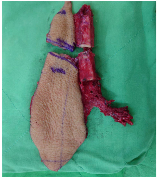
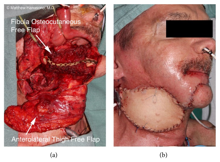
Whatever is chosen, the lining comes first. A small leak of saliva into the neck causes more harm than an imperfect skin match outside.
10B.7 Setting up the operation
- Two teams. One team resects the tumor and does the neck dissection. The second team raises the fibula flap at the same time. This saves several hours.
- Position. The patient lies supine. A support is placed under the buttock on the side of the flap. The knee is flexed to about 90 degrees and the foot is held by a support, so that the lateral side of the leg faces upward.
- Tourniquet. A thigh tourniquet gives a bloodless field. Note the time it is inflated. It is released before the pedicle is divided.
- Preparation. The whole leg is prepared, including the foot, so that the foot can be checked. A thigh is also prepared, for a skin graft and for a second flap if needed.
- Anesthesia. Keep the patient warm and well filled. Avoid vasoconstrictor infusions where possible. Agree this with the anesthetist at the start.
10B.8 Raising the flap
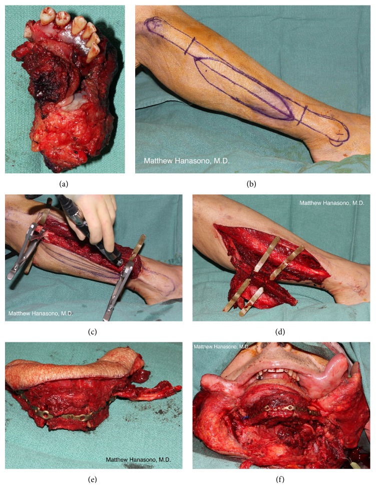
The lateral approach is described here. The dissection goes around the fibula from the lateral side, compartment by compartment, and reaches the pedicle last.
Flowchart: Order of the dissection
- Marking
- Anterior incisionfind the perforators
- Lateral compartmentperoneal muscles
- Anterior compartmentto the interosseous membrane
- Posterior incisionsoleus
- Bone cutslower, then upper
- Interosseous membrane
- Pediclefrom below upward
Step 1. Marking
- Mark the head of the fibula and the lateral malleolus. Draw the line between them along the posterior border of the fibula. This line lies over the posterior crural septum.
- Mark 6 cm below the head of the fibula and 6 to 8 cm above the lateral malleolus. Bone is taken only between these marks.
- Mark the common peroneal nerve at the neck of the fibula.
- Find the perforators with a handheld Doppler probe along the line, mainly in the lower half of the leg.
- Draw the skin paddle as an ellipse centered on the perforators, over the junction of the middle and lower thirds. Draw it larger than the defect. If two paddles are planned, mark a second ellipse over the proximal perforator.
Step 2. Anterior incision and the perforators
- Incise the anterior edge of the skin paddle, through skin, fat and deep fascia, onto the peroneal muscles.
- Lift the skin and fascia off the peroneal muscles, working backward, until the posterior crural septum is seen.
- Look for the perforators in the septum. Confirm that they enter the skin paddle. Adjust the paddle now if the perforators are not where the Doppler suggested.
- If no septal perforator is found, look for perforators through soleus. They can be used if a cuff of soleus is taken with them.
Step 3. Lateral and anterior compartments
- Separate peroneus longus and brevis from the lateral surface of the fibula. Leave a thin layer of muscle (1 to 2 mm) on the bone. Do not strip the periosteum.
- Continue around the front of the bone. Divide the anterior crural septum.
- Separate the extensor muscles (extensor digitorum longus and extensor hallucis longus) from the fibula, again leaving a thin cuff.
- The anterior tibial vessels and deep peroneal nerve lie on the interosseous membrane here. Stay close to the fibula.
- Stop when the interosseous membrane is seen along the whole length of the bone to be taken.
Step 4. Posterior incision
- Incise the posterior edge of the skin paddle through the deep fascia, onto soleus.
- Lift the skin and fascia off soleus, working forward to the septum. The skin paddle now hangs on the septum and its perforators.
- Separate soleus from the back of the fibula and from flexor hallucis longus (FHL). Leave FHL attached to the fibula.
Step 5. Bone cuts
- At each mark, clear a few millimeters of bone all around and pass a curved instrument behind it to protect the vessels.
- Cut the lower end first, then the upper end, with an oscillating saw and irrigation. At the upper cut, the common peroneal nerve is close.
- The fibula can now be pulled outward with a bone hook. This opens the space between the fibula and the tibia.
Step 6. Interosseous membrane and pedicle
- Divide the interosseous membrane along the length of the bone, close to the fibula. The fibers of tibialis posterior appear behind it. They have a V shape.
- At the lower bone cut, find the peroneal vessels behind the fibula. Ligate and divide them there.
- Follow the vessels upward. Divide tibialis posterior along its middle to open the space over the vessels. Divide FHL, leaving a cuff of FHL on the flap. Ligate the muscular branches as they are met.
- Continue up to the origin of the peroneal artery from the tibioperoneal trunk. The posterior tibial vessels and tibial nerve lie just medial. Do not confuse them with the pedicle.
Step 7. Before the pedicle is divided
- Release the tourniquet. Check that the bone ends, the muscle cuff and the skin paddle bleed.
- Check the foot. It should become pink, and the pulses should return. If there is any doubt, place a soft clamp on the peroneal artery and check the foot again before dividing it.
- Leave the flap attached and perfused in the leg. Do the bone shaping now if possible. Divide the pedicle only when the recipient site and vessels are ready.
Points that protect the flap and the leg.
- See the perforators before cutting the whole skin paddle.
- Do not separate the skin paddle from the septum. Do not pull on it.
- Keep the periosteum and a thin muscle cuff on the bone.
- Protect the common peroneal nerve at the upper end and the superficial peroneal nerve in the lateral compartment.
- Keep 6 to 8 cm of fibula above the ankle.
- Check the foot before dividing the pedicle.
10B.9 Shaping the bone
10B.9.1 Principles
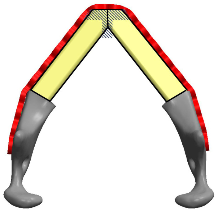
The fibula is straight. The mandible is curved. The fibula is cut into segments so that it follows the curve. Each cut is a closing wedge osteotomy: a small wedge of bone is removed and the two ends are brought together.
- Shape the bone while it is still perfused in the leg. This shortens the time the flap is without blood (ischemia time).
- Do not strip the periosteum. At each cut, open the periosteum only along the line of the saw and lift it for a few millimeters.
- Cut from the lateral side. The pedicle lies on the opposite side. Place a flat retractor between the bone and the pedicle before each cut.
- Irrigate. Heat from the saw kills bone.
- Do not make the segments too short. Each segment needs enough periosteal branches to survive. Segments shorter than about 2 to 3 cm are at risk.
- The cut ends must touch. A gap of 1 mm or more at a junction greatly increases the risk of nonunion. On follow-up CT, 79% of junctions with a gap of 1 mm or more did not unite completely, compared with 8% of junctions in contact (Swendseid 2020).
| Defect | Fibula segments | Osteotomies in the fibula | Junctions that must unite |
|---|---|---|---|
| Lateral (body only) | 1 | 0 | 2 |
| Angle to midline | 2 | 1 | 3 |
| Angle to angle, across the chin | 3 | 2 | 4 |
10B.9.2 The plate
| One reconstruction plate | Several miniplates | |
|---|---|---|
| What it is | One long, thick locking plate (2.0 to 2.5 mm system) across the whole reconstruction | One or two small plates (2.0 mm system) across each junction |
| Advantages | Strong. Holds the shape of the whole arch. Can be bent before the resection, so the position of the condyles and the bite are kept. | Thin, less bulky under the skin and mucosa. Easy to adjust each junction. Easier to remove before dental implants. |
| Disadvantages | Bulky; more likely to show or become exposed under thin or irradiated skin. Carries more of the load, so the bone is loaded less. | Less rigid. The shape of the arch must be controlled in another way while the plates are applied. |
Clinical studies have not shown a clear difference. In 143 flaps, complication rates were 27% with miniplates and 30% with reconstruction plates (Shaw 2004). In 117 fibula flaps, nonunion was 14% with miniplates and 13% with reconstruction plates (Robey 2008). Bone contact matters more than the type of plate.
Applying the plate:
- The plate lies on the lateral surface of the fibula, away from the pedicle.
- Use at least two screws in each fibula segment. Screws in the fibula usually engage only the near cortex, so that they do not reach the pedicle on the far side.
- Use three or four screws in the native mandible on each side.
- Locking screws are preferred. The plate then does not need to be pressed onto the bone, which spares the periosteum.
- Place screws away from the osteotomy lines and away from where dental implants are planned.
10B.9.3 Height, condyle and teeth
- Height. A single fibula is about half the height of a mandible with teeth. If it is placed at the lower border, the contour of the jaw is good but the bone is far below the opposing teeth. If it is placed higher, it suits dental implants better but the lower border is less defined. A double-barrel fibula, folded into two parallel segments on one pedicle, gives both (Jones 1988). It needs more bone length and is used mainly for defects of the body.
- Condyle. If the condyle is kept, the fibula is plated to it. If it is removed, the end of the fibula is rounded and placed in the joint socket, or a prosthetic condyle is used.
- Dental implants. The fibula holds dental implants well. They can be placed at the first operation or later.
10B.10 Inset
Inset means placing and fixing the flap in the defect. The order matters, because each step makes the next one harder to reach.
Flowchart: Order of the inset
- 1. Check the bite and the condyles
- 2. Fix the boneplate to the native mandible
- 3. Close the liningfrom the back forward, watertight
- 4. Join the vesselsartery and one or two veins
- 5. Fill dead spacemuscle cuff
- 6. Close the outer skinsecond paddle or second flap
Bone
- Bring the remaining teeth into the correct bite and hold them there, with intermaxillary fixation or by returning a pre-bent plate to its pre-drilled holes.
- Check that both condyles are seated in their sockets. A condyle fixed out of its socket gives an open bite and pain later.
- Fix the plate to the native mandible on both sides. Check each junction for contact. Fill any small gap with bone from the removed wedges.
- Check that the pedicle lies without a twist or kink and reaches the recipient vessels.
Lining
- Suture the inner skin paddle to the mucosa, starting at the back, where access is hardest.
- The closure must be watertight. Use the skin paddle to cover the junctions of the bone and the plate.
- The paddle hangs on its septum. Do not stretch it or fold the septum sharply.
Vessels
| Usual choice | Alternatives | |
|---|---|---|
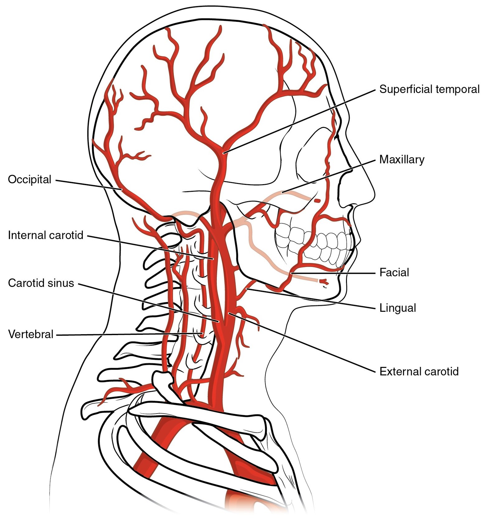
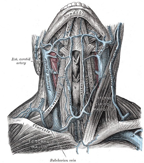
| Artery | Facial artery, end-to-end | Superior thyroid artery, lingual artery, or the external carotid artery end-to-side. In a neck with previous surgery or radiotherapy: the transverse cervical artery, or vessels on the other side of the neck with a vein graft. |
| Vein | Common facial vein or another branch of the internal jugular vein, end-to-end; or the internal jugular vein end-to-side | External jugular vein. Two veins are joined when possible, preferably to two different drainage systems. |
Anatomy of the recipient vessels. The external carotid artery gives branches to the face and neck. From below upward, the three used most are the superior thyroid, the lingual and the facial arteries. The veins lie in two layers: the external and anterior jugular veins under the skin and platysma, and the internal jugular vein with its branches deep to the sternocleidomastoid muscle.
- The time without blood supply should be as short as possible. Shaping in the leg and fixing the bone efficiently are what shorten it.
- After the clamps are released, check bleeding from the skin edge, the muscle cuff and the bone.
- Check the pedicle again with the head in the neutral position and turned to each side.
Dead space and outer skin
- Use the cuff of flexor hallucis longus to fill the space under the jaw and to cover the plate.
- Cover the outer defect with the second paddle or the second flap, without tension.
- Place suction drains in the neck, away from the pedicle and the anastomosis.
- Close the neck without pressure on the pedicle. Do not allow tracheostomy tapes around the neck; suture the tracheostomy tube in place.
10B.11 Closing the leg
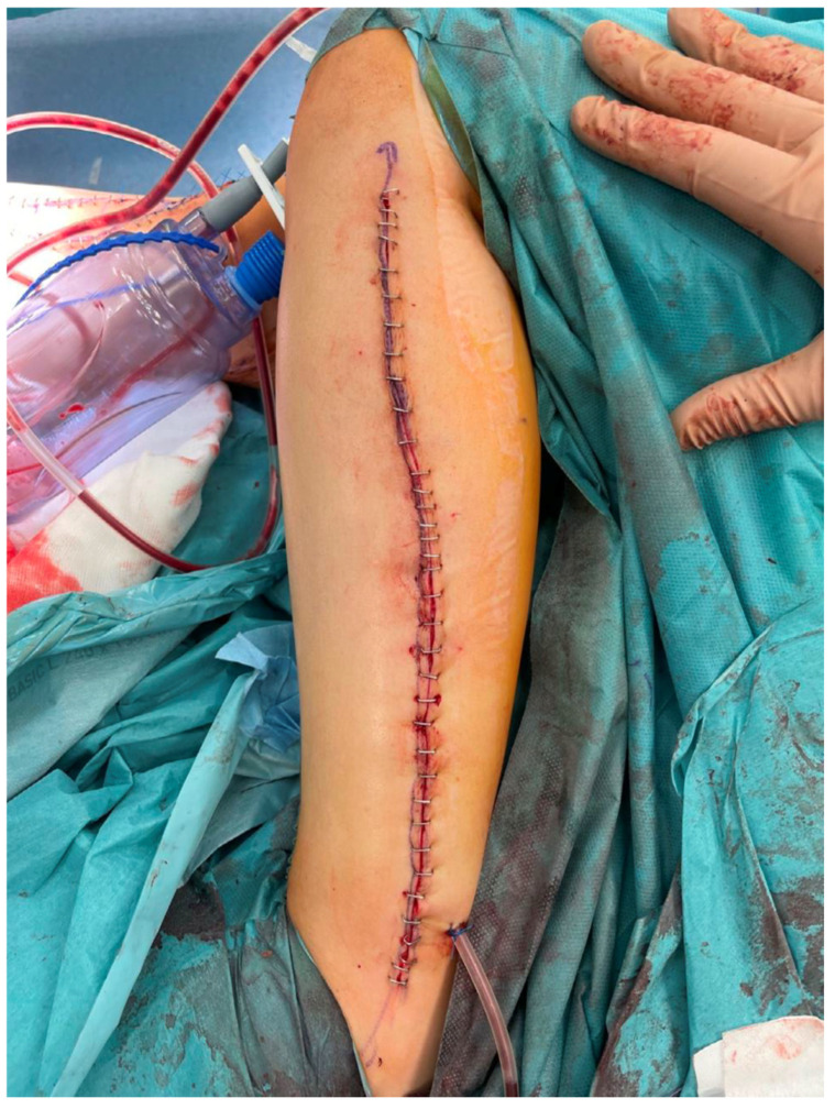
- Secure hemostasis after the tourniquet is released. Place a suction drain.
- The cut end of flexor hallucis longus can be sutured to tibialis posterior or the interosseous membrane. The muscles are laid back loosely. Do not close the deep fascia.
- Skin. If the skin paddle was narrow (about 4 cm or less), the skin can usually be closed directly. If it was wider, use a split skin graft on the muscle. Never close the leg under tension. A tight closure can cause compartment syndrome, skin necrosis and exposed tendons.
- Apply a padded back slab with the ankle at 90 degrees, to protect the skin graft and prevent an equinus position of the foot.
10B.12 Care after surgery
10B.12.1 Monitoring the flap
The bone is buried and cannot be seen. The skin paddle is the monitor for the whole flap.
| Sign | Normal | Arterial problem | Venous problem |
|---|---|---|---|
| Color | Pink, same as the leg | Pale | Blue or purple, mottled |
| Capillary refill | About 2 seconds | Slow or absent | Very fast |
| Turgor | Soft | Flat, empty | Swollen, tense |
| Temperature | Warm | Cool | Cool later |
| Bleeding on needle prick | Bright red, after 1 to 2 seconds | None, or serum only | Dark blood, immediately and briskly |
| Doppler signal | Present | Absent | Arterial signal may remain at first |
- The flap is checked every hour for the first 24 to 48 hours, then less often. Most vascular problems occur in the first two to three days.
- The value of a visible skin paddle is proven. In 750 free flaps, 77% of compromised flaps with a visible part were saved, and none of the buried flaps were saved, because their failure was recognized late (Disa 1999).
- At USM, the skin paddle detected all 4 anastomotic failures early in 13 fibula flaps (Imran 2004).
- The skin paddle is not a perfect monitor. The skin can fail alone, from a problem with its perforator, while the bone is alive. The reverse is rare.
A flap that changes is taken back to the operating room at once. Do not wait to see if it improves. In 1,193 free flaps, flaps that were saved had been re-explored at a mean of 4 hours after the problem was noticed. Flaps that were lost had been re-explored at a mean of 9 hours (Bui 2007).
10B.12.2 General care
- Head position: neutral or turned slightly toward the side of the pedicle, as decided at the end of the operation. No pressure and no ties on the neck.
- Circulation: keep the patient warm, well filled and free of pain. Avoid hypotension.
- Airway: tracheostomy care until the swelling settles.
- Feeding: by nasogastric tube at first. Oral intake starts when the lining has healed, usually after one to two weeks, and later in an irradiated mouth.
- Mouth care: regular gentle cleaning.
- Leg: elevation. Check the color, sensation and movement of the toes. Walking starts after a few days when the wound allows, with physiotherapy. In a prospective study the timing of walking did not affect donor site complications (Momoh 2011).
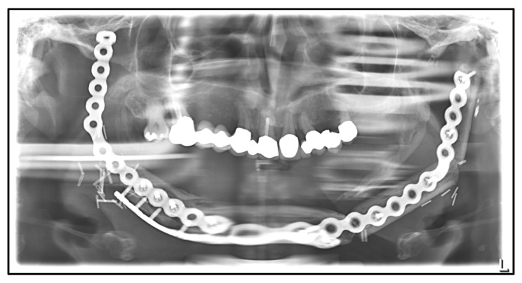
- Imaging: a panoramic radiograph or CT after surgery records the position of the bone and plate. Union is assessed later.
- Radiotherapy, if needed, is planned to start about six weeks after surgery. Wound problems must be solved before then.
10B.13 Complications
Flowchart: Complications of free fibula reconstruction of the mandible
- Complications
- Flapearly
- Venous or arterial thrombosis
Hematoma
Loss of the skin paddle
Total flap loss
- Venous or arterial thrombosis
- Recipient siteearly
- Wound breakdown
Salivary fistula
Infection
Airway problems
- Wound breakdown
- Recipient sitelate
- Plate exposure
Nonunion
Plate fracture
Malocclusion
Osteoradionecrosis
- Plate exposure
- Leg
- Skin graft loss
Ankle stiffness or weakness
Great toe contracture
Nerve injury
Foot ischemia (rare)
- Skin graft loss
- Flapearly
10B.13.1 Problems with the vessels
These are the most urgent complications.
- In 1,193 free flaps, 6% needed emergency re-exploration. The causes were thrombosis of the pedicle in 53% and hematoma or bleeding in 30%. Overall flap survival was 98.8% (Bui 2007).
- Venous thrombosis was more common than arterial thrombosis (74% and 26% of thromboses). It was also saved more often (71% and 40%).
- In 210 bone flaps for the mandible, 16 were re-explored and 8 of these were saved (Urken 1998).
| Cause | How it is prevented |
|---|---|
| Kink or twist of the pedicle | Check the lie of the pedicle after the bone is fixed and again with the head turned |
| Pressure on the pedicle: hematoma, tight closure, tracheostomy tapes | Hemostasis, drains, loose closure, no ties around the neck |
| Poor recipient vessels | Choose vessels outside the irradiated or scarred field when possible; join two veins |
| Technical fault at the anastomosis | Careful microsurgical technique; vessels of matching size |
| Low blood pressure, cold, vasoconstrictors | Agreed plan with the anesthetist and the ward |
Flowchart: Management of a flap that changes
- Change in color, refill, turgor or Doppler signal
- At the bedsiderelease tight sutures and dressings; correct head position; evacuate obvious hematoma
- Return to the operating room without delay
- Explore the pediclerelieve kink or pressure; remove thrombus; redo the anastomosis; vein graft or new recipient vessel if needed
If the skin paddle fails but the bone is alive, the dead skin is removed and replaced with another flap, because the lining must stay sealed. If the whole flap is lost, the dead bone is removed. The options are then a second bone flap, usually the other fibula, or a reconstruction plate covered by a pedicled flap such as pectoralis major, with bone reconstruction later.
10B.13.2 Wound, fistula and infection
- A leak at the lining lets saliva into the neck. It presents with redness, swelling, fever or discharge from the neck wound, usually in the first two weeks.
- Saliva around the anastomosis can cause thrombosis or bleeding from the vessels. A fistula near the pedicle is treated actively: drainage away from the vessels, antibiotics, no oral intake, and early repair with fresh tissue if it does not settle.
- Risk factors are previous radiotherapy, poor nutrition, diabetes, tension on the closure and a skin paddle with poor blood supply.
10B.13.3 Plate exposure
- The plate becomes visible through the skin or the mucosa, months to years after surgery.
- In 221 fibula reconstructions with patient-specific plates, the plate was removed in 11% at a mean of 17 months. The reason was exposure in 76% and loose screws in 24% (Yang 2024).
- Causes: thin or irradiated soft tissue over a bulky plate, wound breakdown, infection, and loose fixation over a junction that has not united.
- Prevention: enough well-vascularized soft tissue over the plate, a low-profile plate where the cover is thin, and no tension on the closure.
- Treatment: if the bone has united, the plate is removed. If it has not, the cause is treated: the nonunion is fixed and grafted, and fresh soft tissue is brought in.
10B.13.4 Nonunion and plate fracture
- Most junctions unite: 97% of osteotomy sites in 150 patients (Cordeiro 1999).
- The causes of nonunion are the ones that apply to any bone: a gap, movement, poor blood supply, infection, and radiotherapy. In this operation the specific causes are poor contact at a junction, a segment that is too short or has lost its periosteum, and a leak of saliva onto the junction.
- The junction between the fibula and the native mandible fails more often than a junction between two fibula segments, because the native bone may be irradiated and the two bones differ in shape.
- A plate across a junction that never unites is loaded at every bite. It will eventually break or its screws will loosen. A broken plate means a nonunion until proven otherwise.
- Treatment follows the general rules: remove dead and infected tissue, restore bone contact or add bone graft, fix rigidly, and make sure the soft tissue cover is healthy.
10B.13.5 Other late problems at the recipient site
| Problem | Cause | Prevention or treatment |
|---|---|---|
| Malocclusion; pain at the joint | Condyle not seated, or bone fixed with the bite not held | Hold the bite and check both condyles before fixing. Late correction needs an osteotomy. |
| Poor contour; visible step | Fibula placed too far in or out; low height; loss of soft tissue after radiotherapy | Accurate planning; fat grafting or contour surgery later |
| Trismus (limited mouth opening) | Scar and fibrosis, radiotherapy | Early mouth-opening exercises |
| Bulky skin paddle in the mouth; no sulcus for a denture | Skin is thicker than mucosa and does not attach to bone | Thinning of the paddle and sulcus surgery later, often with implant surgery |
| Osteoradionecrosis of the native mandible or the fibula | Radiotherapy | Good soft tissue cover; dental care before radiotherapy; avoid surgery in irradiated bone where possible |
| Tumor recurrence | The disease | Follow-up. The plate and flap make imaging more difficult. |
10B.13.6 The leg
Problems in the leg are common early and uncommon late.
| Study | Early problems | Late problems |
|---|---|---|
| Momoh 2011: 157 consecutive patients, prospective | 31% had an early complication: skin graft loss 15%, cellulitis 10%, wound dehiscence 8%, abscess 1% | 17% had a late problem: decreased ankle movement 12%, great toe contracture 9%, leg weakness 8%, ankle instability 4%. 96% returned to their previous level of walking. |
| Ling and Peng 2012: systematic review of 42 studies | Delayed wound healing 17%. Early wound problems in 10% after direct closure and 19% after skin graft. | Limited ankle movement 12%, sensory loss 7%, chronic pain 7%, claw toe 6%, ankle instability 6%, significant gait change 4% |
- Great toe contracture (a flexed great toe) follows scarring or ischemia of flexor hallucis longus. Gentle handling of the muscle, avoiding a tight closure and early toe movement reduce it.
- Nerve injury. Common peroneal nerve injury causes foot drop; the nerve is at risk at the upper bone cut and from retractors. Superficial peroneal nerve injury causes numbness over the top of the foot.
- Compartment syndrome follows a tight skin closure or a hematoma. Pain out of proportion and pain on moving the toes are the signs. The wound is opened at once.
- Ankle instability is prevented by keeping 6 to 8 cm of fibula above the ankle. In children the ankle may drift into valgus with growth, so the lower fibula is fixed to the tibia with a screw in young children.
- Foot ischemia is rare and serious. It is prevented by assessment before surgery and by checking the foot before the pedicle is divided.
10B.14 Function and dental rehabilitation
The purpose of the operation is function: eating, speaking and appearance.
- In 150 patients, 45% returned to an unrestricted diet and 45% to a soft diet. Speech was normal or near normal in 63%. Appearance was judged excellent or good in 59% (Cordeiro 1999).
- Function depends as much on the soft tissue that was removed (tongue, lip, nerves) as on the bone.
- Dental implants. In 210 mandibular reconstructions, 360 implants were placed in 81 patients. Overall implant success was 92%. It was 86% when the bone was irradiated after the implants were placed, and 64% when implants were placed into bone that had already been irradiated (Urken 1998).
- For this reason, when radiotherapy is expected, some units place the implants at the first operation.
10B.15 USM experience
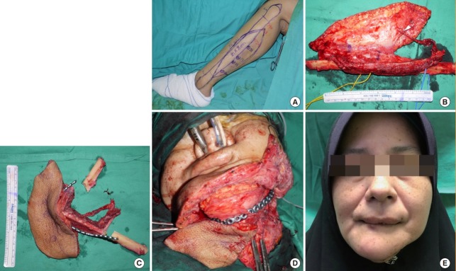
Chai and colleagues reported 27 patients treated at USM over 20 years (1997 to 2017) for ameloblastoma of the mandible. All had radical resection and immediate reconstruction with a free fibula osteocutaneous flap. Flap survival was 100%. Two patients needed re-exploration for venous thrombosis. There was no tumor recurrence at a mean follow-up of 5.6 years (Chai 2019).
10B.16 Summary
- A segmental defect of the mandible, especially an anterior one, needs vascularized bone. A plate with soft tissue alone fails often.
- The fibula gives long, strong bone that can be cut into segments, because each segment lives on its periosteal supply. The periosteum and a thin muscle cuff are always kept.
- The pedicle is the peroneal artery and its veins. The skin is supplied by perforators in the posterior crural septum. See the perforators before committing the skin paddle.
- Before surgery, confirm that the foot does not depend on the peroneal artery.
- When the skin of the face is also removed, decide before the harvest how lining and skin will both be covered: one folded paddle, two paddles, or a second flap.
- Shape the bone while it is perfused. The cut ends must touch. Fix the bone with the bite held and the condyles seated.
- Close the lining first and make it watertight. Then join the vessels.
- The skin paddle is the monitor. A flap that changes goes back to the operating room at once.
- The main late problems are plate exposure and nonunion. Both are prevented by bone contact, stable fixation and healthy soft tissue cover.
References
- Hidalgo DA. Fibula free flap: a new method of mandible reconstruction. Plast Reconstr Surg 1989;84:71–79.
- Brown JS, Lowe D, Kanatas A, Schache A. Mandibular reconstruction with vascularised bone flaps: a systematic review over 25 years. Br J Oral Maxillofac Surg 2017;55:113–126.
- Brown JS, Barry C, Ho M, Shaw R. A new classification for mandibular defects after oncological resection. Lancet Oncol 2016;17:e23–e30.
- Jewer DD, Boyd JB, Manktelow RT, et al. Orofacial and mandibular reconstruction with the iliac crest free flap: a review of 60 cases and a new method of classification. Plast Reconstr Surg 1989;84:391–403.
- Boyd JB, Mulholland RS, Davidson J, Gullane PJ, et al. The free flap and plate in oromandibular reconstruction: long-term review and indications. Plast Reconstr Surg 1995;95:1018–1028.
- Wei FC, Celik N, Yang WG, Chen IH, et al. Complications after reconstruction by plate and soft-tissue free flap in composite mandibular defects and secondary salvage reconstruction with osteocutaneous flap. Plast Reconstr Surg 2003;112:37–42.
- Cordeiro PG, Disa JJ, Hidalgo DA, Hu QY. Reconstruction of the mandible with osseous free flaps: a 10-year experience with 150 consecutive patients. Plast Reconstr Surg 1999;104:1314–1320.
- Gholami M, Hedjazi A, Kiamarz Milani A. Evaluation of anatomic variations of fibula free flap in human fresh cadavers. World J Plast Surg 2019;8:229–236.
- Yu P, Chang EI, Hanasono MM. Design of a reliable skin paddle for the fibula osteocutaneous flap: perforator anatomy revisited. Plast Reconstr Surg 2011;128:440–446.
- Schusterman MA, Reece GP, Miller MJ, Harris S. The osteocutaneous free fibula flap: is the skin paddle reliable? Plast Reconstr Surg 1992;90:787–793.
- Wei FC, Seah CS, Tsai YC, Liu SJ, et al. Fibula osteoseptocutaneous flap for reconstruction of composite mandibular defects. Plast Reconstr Surg 1994;93:294–304.
- Wei FC, Chuang SS, Yim KK. The sensate fibula osteoseptocutaneous flap: a preliminary report. Br J Plast Surg 1994;47:544–547.
- Lutz BS, Wei FC, Ng SH, Chen IH, et al. Routine donor leg angiography before vascularized free fibula transplantation is not necessary: a prospective study in 120 clinical cases. Plast Reconstr Surg 1999;103:121–127.
- Garvey PB, Chang EI, Selber JC, Skoracki RJ, et al. A prospective study of preoperative computed tomographic angiographic mapping of free fibula osteocutaneous flaps for head and neck reconstruction. Plast Reconstr Surg 2012;130:541e–549e.
- Hanasono MM, Skoracki RJ. Computer-assisted design and rapid prototype modeling in microvascular mandible reconstruction. Laryngoscope 2013;123:597–604.
- Tang NSJ, Ahmadi I, Ramakrishnan A. Virtual surgical planning in fibula free flap head and neck reconstruction: a systematic review and meta-analysis. J Plast Reconstr Aesthet Surg 2019;72:1465–1477.
- Rendenbach C, Steffen C, Hanken H, Schluermann K, et al. Complication rates and clinical outcomes of osseous free flaps: a retrospective comparison of CAD/CAM versus conventional fixation in 128 patients. Int J Oral Maxillofac Surg 2019;48:1156–1162.
- Knitschke M, Sonnabend S, Roller FC, Pons-Kühnemann J, et al. Osseous union after mandible reconstruction with fibula free flap using manually bent plates vs. patient-specific implants: a retrospective analysis of 89 patients. Curr Oncol 2022;29:3375–3392.
- Lindhardt JL, Kiil BJ, Jakobsen AM, Buhl J, et al. Implementation of in-house computer-aided design and manufacturing for accelerated free fibula flap reconstruction of mandibular defects in cancer patients. Plast Reconstr Surg Glob Open 2024;12:e6108.
- Zweifel DF, Simon C, Hoarau R, Pasche P, et al. Are virtual planning and guided surgery for head and neck reconstruction economically viable? J Oral Maxillofac Surg 2015;73:170–175.
- Ko YY, Su YX, Pu JJ. Application of temporary patient-specific plate in head and neck osteocutaneous free flap reconstruction: a prospective pilot study. Front Oral Health 2026;7:1692401.
- Lentge F, Jehn P, Neuhaus MT, Bettag SA, et al. A novel method for secondary mandible reconstruction to re-achieve a native condyle position comprising a new design for cutting guides and new positioning devices. J Pers Med 2024;14:181.
- Hanasono MM. Reconstructive surgery for head and neck cancer patients. Adv Med 2014;2014:795483.
- Yagi S, Kamei Y, Torii S. Donor side selection in mandibular reconstruction using a free fibular osteocutaneous flap. Ann Plast Surg 2006;56:622–627.
- Yang KC, Leung JK, Chen JS. Double-paddle peroneal tissue transfer for oromandibular reconstruction. Plast Reconstr Surg 2000;106:47–55.
- Wei FC, Demirkan F, Chen HC, Chen IH. Double free flaps in reconstruction of extensive composite mandibular defects in head and neck cancer. Plast Reconstr Surg 1999;103:39–47.
- Chen HC, Demirkan F, Wei FC, Cheng SL, et al. Free fibula osteoseptocutaneous-pedicled pectoralis major myocutaneous flap combination in reconstruction of extensive composite mandibular defects. Plast Reconstr Surg 1999;103:839–845.
- Chen CC, Chiu TH, Mohammed AA, Shih HS, et al. Adjustable single-osteotomy fibular free flap for anterior mandibular defects in irradiated head and neck cancers: a case series. J Clin Med 2025;14:1953.
- Rodriguez IE, Trinh BB, Deleyiannis FW. Utility of an anterior tibial perforator for skin paddle viability in through-and-through defects of the mandible. Eplasty 2018;18:e24.
- Fouaad AA, Massoud AH, Shaban M, Abdel-Naby M, et al. Vascularized versus nonvascularized free fibular grafts in reconstruction of post-traumatic critical long bone defects, a comparative study. J Orthop Traumatol 2026;27:1.
- Swendseid B, Kumar A, Sweeny L, et al. Natural history and consequences of nonunion in mandibular and maxillary free flaps. Otolaryngol Head Neck Surg 2020;163:956–962.
- Shaw RJ, Kanatas AN, Lowe D, Brown JS, et al. Comparison of miniplates and reconstruction plates in mandibular reconstruction. Head Neck 2004;26:456–463.
- Robey AB, Spann ML, McAuliff TM, et al. Comparison of miniplates and reconstruction plates in fibular flap reconstruction of the mandible. Plast Reconstr Surg 2008;122:1733–1738.
- Jones NF, Swartz WM, Mears DC, Jupiter JB, et al. The "double barrel" free vascularized fibular bone graft. Plast Reconstr Surg 1988;81:378–385.
- Disa JJ, Cordeiro PG, Hidalgo DA. Efficacy of conventional monitoring techniques in free tissue transfer: an 11-year experience in 750 consecutive cases. Plast Reconstr Surg 1999;104:97–101.
- Imran Y, Zulmi W, Halim AS. Skin paddle as an indicator of the viability of vascularised fibular graft. Singapore Med J 2004;45:110–112.
- Bui DT, Cordeiro PG, Hu QY, Disa JJ, et al. Free flap reexploration: indications, treatment, and outcomes in 1193 free flaps. Plast Reconstr Surg 2007;119:2092–2100.
- Urken ML, Buchbinder D, Costantino PD, Sinha U, et al. Oromandibular reconstruction using microvascular composite flaps: report of 210 cases. Arch Otolaryngol Head Neck Surg 1998;124:46–55.
- Yang S, Morton Z, Sautter M, Young G, et al. Long-term plate complications in patient-specific plates utilizing computer-aided design. Laryngoscope 2024;134:4929–4934.
- Kasper R, Scheurer M, Pietzka S, Sakkas A, et al. MRONJ of the mandible: from decortication to a complex jaw reconstruction using a CAD/CAM-guided bilateral scapula flap. Medicina (Kaunas) 2023;59:535.
- Momoh AO, Yu P, Skoracki RJ, Liu S, et al. A prospective cohort study of fibula free flap donor-site morbidity in 157 consecutive patients. Plast Reconstr Surg 2011;128:714–720.
- Ling XF, Peng X. What is the price to pay for a free fibula flap? A systematic review of donor-site morbidity following free fibula flap surgery. Plast Reconstr Surg 2012;129:657–674.
- Opyrchał J, Pachuta B, Bula D, Dowgierd K, et al. Upper limb-salvage surgery in pediatric patients with malignant bone tumors using microsurgical free flaps: long-term follow-up. Biomedicines 2025;13:1638.
- Chai KS, Omar FH, Mat Saad AZ, Wan Sulaiman WA, Halim AS. A 20-year experience of immediate mandibular reconstruction using free fibula osteocutaneous flaps following ameloblastoma resection. Arch Plast Surg 2019;46:426–432.