8.1 Introduction
A bone graft is bone that is moved from one place to another to help a bone defect heal. The word is also used more loosely for any material placed in a bone defect for the same purpose.
Bone healing needs four things, in a bed with a good blood supply: stability, cells, growth factors and a scaffold. This is called the diamond concept (Giannoudis 2007). When a defect does not heal by itself, one or more of these is missing. A graft is chosen to supply what is missing.
Example: the alveolar cleft. A child with a repaired cleft lip and palate has a gap in the alveolar bone. The gap is stable and has a good blood supply, but it never fills with bone by itself. What is missing is bone-forming cells and a scaffold. Boyne and Sands (1972) filled the gap with cancellous bone from the iliac crest, and the canine tooth erupted through the graft. This example is used again in this chapter.
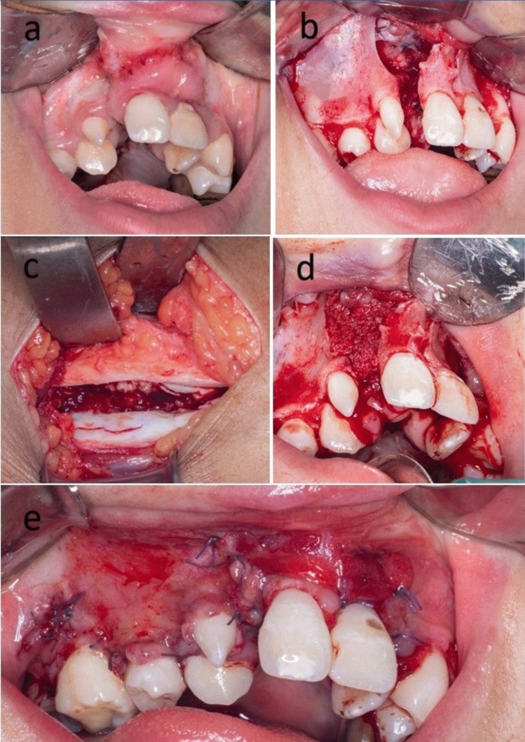
The chapter follows the same order as the decision: what a graft must supply, which material supplies it, what happens after it is placed, and what decides whether it succeeds. These principles apply to every graft material.
Flowchart: Overview of bone graft biology
- What bone healing needs (diamond concept)
- Stabilityfixation
- Cells
- Osteogenesisautograft, vascularized bone, marrow
- Growth factors
- Osteoinductionautograft, demineralized bone matrix, BMP
- Scaffold
- Osteoconductionalmost all grafts and substitutes
- Blood supplythe bed, or a flap
8.2 What a graft must supply
A graft is chosen for what it brings to the defect. This is described by four properties. They mean different things and should not be mixed up.
| Term | Meaning | Which materials have it |
|---|---|---|
| Osteogenesis | The graft contains living cells that make new bone. | Only fresh autograft, marrow and vascularized bone |
| Osteoinduction | The graft contains signals (growth factors) that make the patient's own stem cells turn into bone-forming cells. | Autograft, demineralized bone matrix, BMP |
| Osteoconduction | The graft is a scaffold. Vessels and new bone grow along its surface and into its pores. | Almost all grafts and bone substitutes |
| Osseointegration | Bone attaches directly to an implant, with no fibrous tissue in between. | Titanium implants |
- Osteoinduction was discovered by Urist (1965). He put demineralized bone into muscle, and new bone formed there. The active substances were later named bone morphogenetic proteins.
- Osseointegration was described by Brånemark, using titanium implants in the jaw (1977).
- A fifth property is often added: mechanical support. A cortical graft or a block can carry load from the first day. A cancellous graft cannot.
In the alveolar cleft example, cancellous autograft works well because it has the first three properties together.
The osteoinductive signals in bone matrix are the bone morphogenetic proteins (BMP).
8.3 Which material supplies what
Graft materials are grouped by where they come from and by the type of bone. Each group supplies a different set of the four properties.
8.3.1 Classes of graft
A graft is described in several ways at once. For example, "a non-vascularized cortical autograft from the calvarium, used as an onlay".
| Described by | Types | Meaning |
|---|---|---|
| Source | Autograft | From the same patient |
| Allograft | From another person, usually a deceased donor, through a bone bank | |
| Xenograft | From another species, usually processed bovine bone | |
| Alloplast (bone substitute) | Synthetic material. Strictly it is an implant and not a graft. | |
| Tissue | Cancellous | Trabecular bone with marrow; many cells, no strength |
| Cortical | Dense bone; few cells, strong | |
| Corticocancellous | A block with both, for example a piece of iliac crest | |
| Blood supply | Non-vascularized (free graft) | Moved without vessels; depends on the bed |
| Vascularized | Moved with its artery and vein, as a pedicled or free flap | |
| Position | Inlay | Placed inside a bone defect, with bone on its sides |
| Onlay | Placed on the surface of a bone, to add contour | |
| Interpositional | Placed in the gap between two bone ends, for example after an osteotomy | |
| Origin of the bone | Membranous | From bone formed by intramembranous ossification: calvarium, facial bones |
| Endochondral | From bone formed through cartilage: iliac crest, rib, tibia |
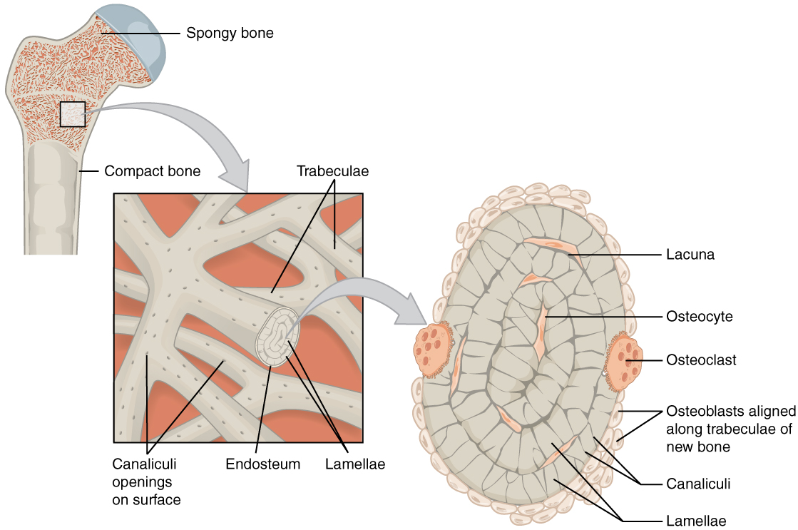
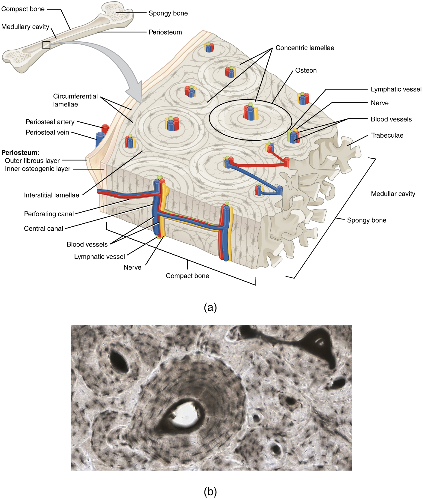
This difference in structure explains most of the difference in behavior between cancellous and cortical grafts.
8.3.2 Comparing what each class supplies
The four properties allow all the materials to be compared. Only vascularized bone has all of them. Most bone substitutes have only one.
| Material | Osteogenic (cells) | Osteoinductive (signal) | Osteoconductive (scaffold) | Strength at first | Own blood supply |
|---|---|---|---|---|---|
| Cancellous autograft | +++ | ++ | +++ | – | – |
| Cortical autograft | + | + | + | +++ | – |
| Vascularized bone flap | +++ | ++ | +++ | +++ | +++ |
| Bone marrow aspirate | ++ | + | – | – | – |
| Structural allograft (frozen) | – | + | + | +++ | – |
| Demineralized bone matrix | – | ++ | + | – | – |
| Xenograft (bovine bone mineral) | – | – | ++ | + | – |
| Calcium phosphate ceramics | – | – | ++ | + | – |
| rhBMP-2 on collagen sponge | – | +++ | – | – | – |
| Titanium, PEEK, PMMA | – | – | – | +++ | – |
Grades are the usual textbook grades. (Burchardt 1983; Bauer and Muschler 2000; Albrektsson and Johansson 2001)
Autograft is the standard against which the others are compared. Its limits are the amount available and the harm to the donor site. The other materials were developed to reduce these two limits.
8.3.3 What the surgeon is handed: examples of products
| Class | What the surgeon is handed | Examples of products |
|---|---|---|
| Autograft | The patient's own bone, harvested at the same operation | No product. Harvesting tools include bone scrapers, trephines and the reamer-irrigator-aspirator. |
| Allograft bone | Frozen or freeze-dried donor bone from a tissue bank: chips, blocks, struts, femoral heads | Supplied by tissue banks, including the USM Tissue Bank. Processed brands include Puros and Tutoplast. |
| Demineralized bone matrix | Putty, gel or paste in a syringe or jar | Grafton, DBX, Allomatrix |
| Xenograft | Granules or blocks of bovine bone mineral | Bio-Oss, Endobon, cerabone |
| Calcium phosphate ceramics | Granules, blocks, or a cement that is mixed and sets in the defect | Vitoss, chronOS, Cerasorb (β-TCP); Pro Osteon (hydroxyapatite); Norian, HydroSet (cements) |
| Bioactive glass | Granules or putty | NovaBone, BonAlive |
| Growth factor | A vial of protein with a collagen sponge | Infuse (rhBMP-2) |
| Cell and platelet concentrates | A disposable kit and a bench centrifuge | SmartPrep and Angel (marrow); Arthrex ACP, GPS III (PRP) |
| Implants | Titanium mesh, custom PEEK implant, porous polyethylene shapes, bone cement | Medpor (porous polyethylene); Palacos, Simplex P (PMMA cement) |
8.4 What happens after the graft is placed
A vascularized bone flap stays alive and heals like a fracture. A non-vascularized graft must be incorporated. Incorporation is the process by which a graft becomes part of the host bone. The graft is revascularized, united to the host, and gradually replaced by new living bone.
8.4.1 What survives the transfer
A free graft has no blood supply at first. Its cells live by diffusion of fluid from the bed, as in a skin graft.
- Oxygen and nutrients diffuse only about 0.1 to 0.2 mm. Only cells on or near a surface survive.
- Osteocytes deep inside cortical bone die. Their empty lacunae are seen on histology.
- The cells that survive and make the first new bone are surface cells. In cortical bone isografts in the rat, the endosteal lining cells and marrow stroma produced more than half of the new bone, and the periosteal cells about 30%. Osteocytes contributed about 10% (Gray and Elves 1979).
This is why cancellous bone, which is almost all surface, is the most osteogenic graft. It is also why a graft should be kept moist, handled gently and placed quickly.
8.4.2 Phases of incorporation
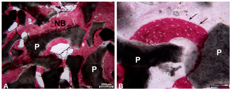
All non-vascularized materials that are replaced by bone (autograft, allograft and resorbable substitutes) pass through the same phases. The phases overlap. The times below are for cancellous autograft in a good bed; the other materials are slower:
Flowchart: Phases of graft incorporation, with approximate times
- Hematoma and inflammationfirst week
- Revascularizationvessels grow in from the bed; from day 2 to 3, complete by about 2 weeks
- Osteoinductionhost stem cells become osteoblasts; from the first to about the fourth week
- Osteoconductionnew bone grows along the graft; from about 2 weeks, for several months
- Remodelingshaped by load; from about 3 months, for months to years
Times are approximate and come mainly from animal studies; they are longer in a poor bed and in older patients (Burchardt 1983).
The replacement of dead graft by new living bone is called creeping substitution. Vessels and cells from the host "creep" into the graft, remove the dead bone and replace it.
Cancellous and cortical bone pass through these phases in a different order. In cancellous bone, new bone is laid on the dead trabeculae first, so the graft becomes stronger from the start and is fully replaced. In cortical bone, osteoclasts must first widen the canals, so the graft becomes weaker for several months (roughly 6 weeks to 6 months) before it recovers, and it is never fully replaced (Enneking 1975).
8.4.3 How each class becomes part of the skeleton
Not every material is incorporated in this way. The table compares all the classes.
| Class | How it becomes part of the host | Speed | Result |
|---|---|---|---|
| Vascularized bone flap | Its cells stay alive. It unites with the host bone in the same way as a fracture heals. There is no creeping substitution. | Like a fracture: weeks to a few months | Living bone that thickens with load. Does not depend on the bed. |
| Cancellous autograft | Revascularized, then new bone is laid on the dead trabeculae, which are later removed | Fast: vessels in about 2 weeks; replaced within about a year | Completely replaced by living bone |
| Cortical autograft | Canals widened by osteoclasts first, then filled with new bone (creeping substitution) | Slow; weakest from about 6 weeks to 6 months | Partly replaced; dead and new bone remain mixed for years |
| Allograft | The same process as autograft, but with no living cells, little signal after processing, and an immune response against donor antigens | Slower than autograft | Often incomplete, especially in large cortical allografts; late fracture and nonunion at the junctions |
| Demineralized bone matrix | Releases BMP, which makes host stem cells form bone (osteoinduction); the collagen then acts as a scaffold | Weeks; depends on the product and batch | Replaced by host bone if the bed is good. No strength. |
| Bovine bone mineral and calcium phosphate ceramics | Scaffold only. New bone grows over the surface and into the pores (osteoconduction). | Depends on the material: calcium sulfate in weeks, β-TCP in months, hydroxyapatite and bovine mineral over years or never | New bone mixed with remaining particles |
| Implants (titanium, PEEK, PMMA, porous polyethylene) | Not incorporated. Titanium osseointegrates: bone attaches directly to its surface. PEEK and PMMA are surrounded by a fibrous capsule. Porous polyethylene is filled by fibrous tissue and vessels. | Not applicable | Permanent foreign body; depends entirely on the soft tissue cover |
8.5 What decides success
A non-vascularized graft or substitute is a passenger. It depends on the bed for its blood supply. Whether it succeeds is decided by the material, the bed, the mechanical conditions, the patient and, finally, the size of the defect. A vascularized flap is much less affected by the bed, which is the main reason for choosing it.
8.5.1 The material, the bed and the mechanics
| Group | Factor | Effect |
|---|---|---|
| The material | Type of bone | Cancellous is revascularized fastest. Cortical gives strength and keeps its volume best. Processed and synthetic materials have no cells. |
| Size and thickness | The center of a large graft is the last part to receive vessels | |
| Handling | Drying, heat from a burr or saw, and a long time out of the body kill the surface cells. Keep the graft in a blood-soaked or saline-soaked swab and use it soon. | |
| The bed | Blood supply | The most important factor. A graft does poorly on scar, on bare cortex, or under thin skin. |
| Previous radiotherapy | The bed has few vessels and few cells. A free graft usually fails; vascularized bone is needed. | |
| Infection or contamination | A free graft has no defense until it is revascularized. Saliva and sinus contents are common causes of loss in the face. | |
| Soft tissue cover | The graft needs well-vascularized cover closed without tension, on every side | |
| Mechanics | Fixation | Movement tears the new vessels. Rigid fixation increases survival. |
| Contact with host bone | Bone forms from the surfaces in contact. Gaps fill with fibrous tissue. | |
| Load | A graft that carries no load is resorbed with time. A graft that is loaded is maintained. | |
| The patient | Smoking, diabetes, malnutrition, steroids, age | The same factors that delay fracture healing |
8.5.2 Why defect size matters
A non-vascularized graft or a scaffold depends on vessels growing in from the surrounding bone. This limits how large a defect it can fill.
- Oxygen can diffuse only about 0.1 to 0.2 mm from a capillary. Cells further away than this die unless new vessels reach them in time.
- For vessels to grow in, the pores of a scaffold must be connected to each other and large enough (about 0.3 mm or more) (Karageorgiou and Kaplan 2005).
- In a large graft, the center is the last part to get a blood supply, and it may die first.
Figure 8.5. Vessels (red) grow into a graft from the host bone on each side. The center (grey) is supplied last. Drawn for this chapter.
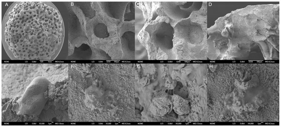
Properties of an ideal scaffold: biocompatible; osteoconductive; interconnected pores (pore size about 100 to 500 µm); resorbs at the same rate as new bone forms; adequate strength; can be shaped; can be sterilized.
8.5.3 Critical-size defect
A critical-size defect is a defect that is too large to heal by itself (Schmitz and Hollinger 1986). The size depends on the bone, the age of the patient and the bed.
| Site | Evidence | Practical limit for a non-vascularized graft |
|---|---|---|
| Long bone | A defect is usually considered critical-sized when it is longer than about 2 cm or involves more than 50% of the circumference (Roddy 2018). | Commonly quoted as about 6 cm for a non-vascularized graft in a good bed |
| Mandible | Non-vascularized grafts unite less often than vascularized flaps, and fail more often as the defect gets longer (Pogrel 1997; Foster 1999). | Non-vascularized grafts for defects shorter than about 5 to 6 cm, in a bed that has not been irradiated. Vascularized bone for longer defects (certainly over 9 cm), irradiated beds, and when soft tissue is also needed. |
| Alveolar cleft | In 93 patients, graft failure rose sharply above a cleft volume of about 0.8 cm³: 71% failure above this size and none below it for iliac crest graft (Roohani 2025). | A wide cleft may need orthodontic narrowing first, or a larger volume of graft with reliable soft tissue closure |
| Skull vault | In infants the dura can close a defect. This ability is lost after about the first year of life. | After infancy, a full-thickness skull defect does not close and needs a graft or an implant |
For large defects there are three solutions: bone that comes with its own blood supply (a vascularized bone flap); new bone grown gradually from the bone ends (distraction osteogenesis); or a vascular bed that is created first and grafted later (a staged technique).
8.6 The alveolar cleft example, explained
The example from the introduction can now be explained step by step.
- What is missing. The cleft is a gap between two bone edges. It is stable and has a good blood supply. What it lacks is bone-forming cells and a scaffold for them.
- Which material. The graft must bring cells and a scaffold. It must also be completely replaced by living bone, because the canine tooth has to erupt through it and later be moved by the orthodontist. Cancellous autograft does all of this. A cortical graft would remain partly dead for years, and bovine bone mineral hardly resorbs, so neither is suitable.
- Position. The graft fills a gap between bone edges, so it is an inlay graft, with bone contact on several sides. Inlay grafts keep their volume well, and the erupting tooth later loads it.
- What decides success. A watertight closure of the nose and mouth around the graft, a graft packed firmly against the bone edges, a cleft that is not too large, and grafting before the canine erupts.
8.7 Summary
- A graft supplies one or more of: cells (osteogenesis), signals (osteoinduction), scaffold (osteoconduction) and strength.
- In a free graft only the surface cells survive. The rest is replaced by creeping substitution.
- Each class becomes part of the skeleton in its own way: a vascularized flap heals like a fracture; autograft and allograft are replaced by creeping substitution; substitutes act as scaffolds; implants are never incorporated.
- Survival depends on the bed, on fixation and on contact, more than on the graft.
- Large defects, and irradiated or scarred beds, need bone that brings its own blood supply.
Before choosing a graft, assess the defect and its bed:
| Question | If the answer is no |
|---|---|
| Is it stable? | Fix it first. No graft survives movement. |
| Does the bed have a good blood supply? | Bring vascularized tissue: a flap, or vascularized bone. |
| Are bone-forming cells present? | Use autograft or vascularized bone. |
| Is the defect small? | If large, a scaffold alone will fail. Use vascularized bone or a staged technique. |
When the bed is good but one part is still missing, a product can sometimes supply that part on its own. These products are called orthobiologics.
References
- Giannoudis PV, Einhorn TA, Marsh D. Fracture healing: the diamond concept. Injury 2007;38 Suppl 4:S3–S6.
- Boyne PJ, Sands NR. Secondary bone grafting of residual alveolar and palatal clefts. J Oral Surg 1972;30:87–92.
- Urist MR. Bone: formation by autoinduction. Science 1965;150:893–899.
- Albrektsson T, Johansson C. Osteoinduction, osteoconduction and osseointegration. Eur Spine J 2001;10 Suppl 2:S96–S101.
- Brånemark PI, Hansson BO, Adell R, et al. Osseointegrated implants in the treatment of the edentulous jaw. Scand J Plast Reconstr Surg Suppl 1977;16:1–132.
- Burchardt H. The biology of bone graft repair. Clin Orthop Relat Res 1983;174:28–42.
- Bauer TW, Muschler GF. Bone graft materials: an overview of the basic science. Clin Orthop Relat Res 2000;371:10–27.
- Gray JC, Elves MW. Early osteogenesis in compact bone isografts: a quantitative study of contributions of the different graft cells. Calcif Tissue Int 1979;29:225–237.
- Enneking WF, Burchardt H, Puhl JJ, Piotrowski G. Physical and biological aspects of repair in dog cortical-bone transplants. J Bone Joint Surg Am 1975;57:237–252.
- Karageorgiou V, Kaplan D. Porosity of 3D biomaterial scaffolds and osteogenesis. Biomaterials 2005;26:5474–5491.
- Schmitz JP, Hollinger JO. The critical size defect as an experimental model for craniomandibulofacial nonunions. Clin Orthop Relat Res 1986;205:299–308.
- Roddy E, DeBaun MR, Daoud-Gray A, Yang YP, Gardner MJ. Treatment of critical-sized bone defects: clinical and tissue engineering perspectives. Eur J Orthop Surg Traumatol 2018;28:351–362.
- Foster RD, Anthony JP, Sharma A, Pogrel MA. Vascularized bone flaps versus nonvascularized bone grafts for mandibular reconstruction: an outcome analysis of primary bony union and endosseous implant success. Head Neck 1999;21:66–71.
- Pogrel MA, Podlesh S, Anthony JP, Alexander J. A comparison of vascularized and nonvascularized bone grafts for reconstruction of mandibular continuity defects. J Oral Maxillofac Surg 1997;55:1200–1206.
- Roohani I, Youn S, Alfeerawi S, Shakoori P, et al. Failure rates based on alveolar cleft volume: analysis of the critical-size defect for alveolar bone grafting. Plast Reconstr Surg 2025;155:377e–386e.
- Giannoudis PV, Dinopoulos H, Tsiridis E. Bone substitutes: an update. Injury 2005;36 Suppl 3:S20–S27.