10.1 Introduction
A non-vascularized bone graft fails in long defects and in poor beds, because it depends on the bed for its blood supply.
A vascularized bone flap is bone transferred together with its artery and vein. The vessels are either kept attached (pedicled flap) or joined to vessels at the recipient site by microsurgery (free flap). The bone stays alive. It does not depend on the bed.
History:
- 1975: Taylor performed the first free vascularized fibula, for a tibial defect.
- 1986: Wei described the skin perforators of the fibula flap, so that skin could be taken reliably with the bone.
- 1989: Hidalgo used the fibula, with osteotomies, to reconstruct the mandible.
Flowchart: Overview of bone transfer
- Autogenous bone transfer
- Non-vascularized graft
- Depends on the bed
- Vascularized bone
- Pedicled
- Calvariumsuperficial temporal
- Distal radiusfor scaphoid
- Free flap
- Fibulaperoneal
- Iliac crestDCIA
- Scapulacircumflex scapular
- Medial femoral condyledescending genicular
- Fibula with epiphysisanterior tibial
- Pedicled
- Non-vascularized graft
10.2 How vascularized bone differs from a bone graft
| Non-vascularized graft | Vascularized bone flap | |
|---|---|---|
| Cells after transfer | Most die | Stay alive |
| How it joins the host bone | Incorporation (creeping substitution), slow | Like a normal fracture, faster |
| Strength | Cortical graft weakens for months | No weak period |
| Depends on the bed | Yes | No: works in irradiated, scarred or infected beds |
| Response to load | Little | Hypertrophy (the bone becomes thicker) |
| Other tissue | Bone only | Can include skin and muscle |
| Disadvantage | Simple operation | Long operation, microsurgery, donor site morbidity |
Hypertrophy and fracture. De Boer and Wood (1989) reviewed 62 vascularized fibula transfers. The fibula became thicker with time, more so when the limb was loaded. But 25% fractured, at a mean of 8 months, before the bone had thickened. So the flap must be protected in the first year, but also loaded gradually so that it hypertrophies.
10.3 Indications
Vascularized bone is indicated when a non-vascularized graft is likely to fail:
- Long segmental defect (commonly quoted as more than 6 cm)
- Poor recipient bed: irradiated, scarred or previously infected
- Composite defect needing bone and soft tissue together
- Nonunion with avascular bone (for example the scaphoid)
- Children who have lost a growth plate
Flowchart: When to use vascularized bone
- Bone defect
- Small defect, healthy bed
- Non-vascularized graft
- Any of these
- Defect over about 6 cm; irradiated, scarred or infected bed; bone + soft tissue needed; avascular nonunion; growth plate lost
- Vascularized bone flap
- Defect over about 6 cm; irradiated, scarred or infected bed; bone + soft tissue needed; avascular nonunion; growth plate lost
- Small defect, healthy bed
10.4 The free fibula flap
The fibula is the most used vascularized bone flap, so it is described first and in most detail.
10.4.1 Anatomy
- Vessel: peroneal artery and its two veins.
- Bone: up to about 25 cm of straight cortical bone. The distal 6 to 8 cm is left in place to keep the ankle stable.
- Blood supply to the bone: one nutrient artery (endosteal supply) and many periosteal branches along its length.
- Skin paddle: supplied by perforators that pass through the posterior crural septum.
More detail:
- The peroneal artery arises from the tibioperoneal trunk and runs down between tibialis posterior and flexor hallucis longus, close to the fibula.
- The nutrient artery enters the middle third of the fibula. The periosteal branches are segmental, which is the basis for multiple osteotomies.
- The pedicle is short at its origin. It is lengthened by removing proximal bone subperiosteally and using the distal fibula for the reconstruction.
- Skin perforators are mostly septocutaneous in the distal and middle thirds of the leg, and more often musculocutaneous (through soleus) proximally. The skin paddle is centered over the posterior border of the fibula at the junction of the middle and distal thirds.
- Proximally, about 6 cm of fibula is preserved to protect the common peroneal nerve and the knee. Distally, 6 to 8 cm is preserved for ankle stability.
- Flexor hallucis longus muscle can be included to fill dead space.
Preoperative assessment: palpate dorsalis pedis and posterior tibial pulses. Imaging (CT or MR angiography) is used when pulses are abnormal, after leg trauma, or with vascular disease. It excludes peronea arteria magna and significant atherosclerosis.
10.4.2 Why the fibula can be osteotomized
When the fibula is cut into segments, the endosteal supply to each segment is interrupted. Each segment survives on its periosteal supply. For this reason:
- the periosteum is never stripped from the fibula, and
- the bone is cut from the side opposite the pedicle.
10.4.3 Shaping the fibula for the mandible
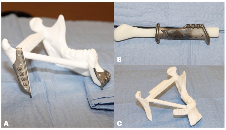
The fibula is straight and the mandible is curved. The fibula is cut into segments (closing wedge osteotomies) to follow the curve. A larger defect needs more osteotomies.
| Defect | Fibula segments | Osteotomies | Junctions that must unite |
|---|---|---|---|
| Lateral (body only) | 1 | 0 | 2 |
| Angle to midline | 2 | 1 | 3 |
| Angle to angle, across the chin | 3 | 2 | 4 |
Every junction must unite. A gap of 1 mm or more at a junction greatly increases the risk of nonunion. The osteotomies must close with good bone contact.
Classification of mandibular defects (Brown 2016). The mandible has four "corners": the two angles and the two canines. The class depends on which corners are removed.
| Class | Defect |
|---|---|
| I | Lateral: includes one angle, not the canine |
| II | Hemimandibulectomy: one angle and the canine on the same side |
| III | Anterior: both canines, neither angle |
| IV | Extensive: both canines and at least one angle |
| "c" | Added if the condyle is also removed (for example class IIc) |
Each corner removed needs one more osteotomy in the fibula.
The older HCL classification (Jewer 1989) is still widely used: H = lateral defect including the condyle; C = central segment including both canines; L = lateral defect without the condyle. Letters are combined (LC, HC, LCL, HCL). Central (C) defects are the most disabling if not reconstructed with bone, because the chin, lip and tongue lose their support ("Andy Gump" deformity).
Goals of mandibular reconstruction: restore continuity; keep the condyles seated and the occlusion correct; restore lower facial height and projection; give enough bone height and width for dental implants; provide intraoral lining and a seal against saliva.
Additional points:
- Height: a single fibula is about half the height of a dentate mandible. Options: place the fibula at the alveolar level, double-barrel the fibula, or later vertical distraction.
- Condyle: if it can be preserved, the fibula is plated to it. If not, the end of the fibula is rounded and placed in the fossa, or a costochondral graft or prosthesis is used.
- Dental implants osseointegrate in fibula. They can be placed at the same operation or later; radiotherapy reduces implant survival.
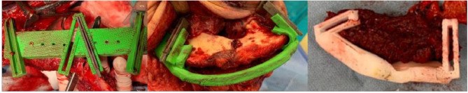
- Virtual surgical planning: CT of the jaw and leg is used to plan the resection and osteotomies. Cutting guides and a pre-bent or custom plate are made. This improves accuracy and shortens ischemia time.
10.4.4 USM experience
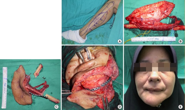
- 27 patients over 20 years (1997 to 2017) with ameloblastoma of the mandible. All had radical resection and immediate free fibula reconstruction. Flap survival was 100%. Two needed re-exploration for venous thrombosis. There was no tumor recurrence at a mean follow-up of 5.6 years.
- the skin paddle is used to monitor the buried bone. In 13 fibula flaps, the skin paddle detected all 4 anastomotic failures early.
- 13 long bone reconstructions with vascularized fibula. Mean time to union was 32 weeks.
10.4.5 Donor site complications
- Ankle stiffness or instability, weakness of great toe flexion.
- In children, the ankle may drift into valgus as the child grows. In a USM review of 8 children, 2 developed ankle valgus; both had surgery before 6 years of age .
- Check the blood supply of the foot before surgery. Rarely the peroneal artery is the main supply to the foot (peronea arteria magna); a USM case of this was reported .
10.5 Other vascularized bone flaps
The fibula is not suitable for every defect. Two things decide the choice of flap: the shape of bone that is needed, and the amount of soft tissue that is needed with it.
10.5.1 The main alternatives to the fibula
| Flap | Vessel | Main use | Main disadvantage |
|---|---|---|---|
| Fibula | Peroneal | Mandible, long bones | Limited height; leg donor site |
| Iliac crest | Deep circumflex iliac (DCIA) | Lateral mandible; good height for implants | Hernia, pain, bulky |
| Scapula | Circumflex scapular; angular branch | Maxilla and palate; defects needing a lot of soft tissue | Patient must be turned; shorter bone |
| Radial forearm with bone | Radial | Small defects needing thin skin | Fracture of the radius |
| Medial femoral condyle | Descending genicular | Scaphoid nonunion, small nonunions | Small amount of bone |
| Calvarium (pedicled) | Superficial temporal | Orbit and midface | Limited size and reach |
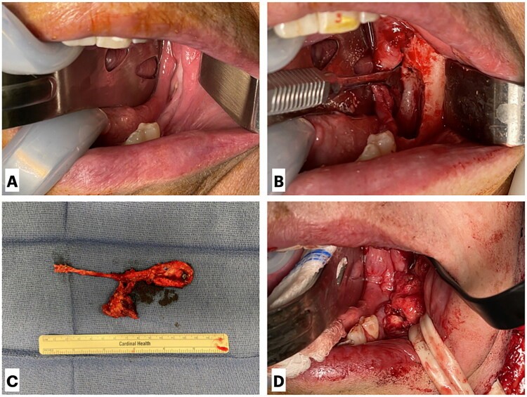
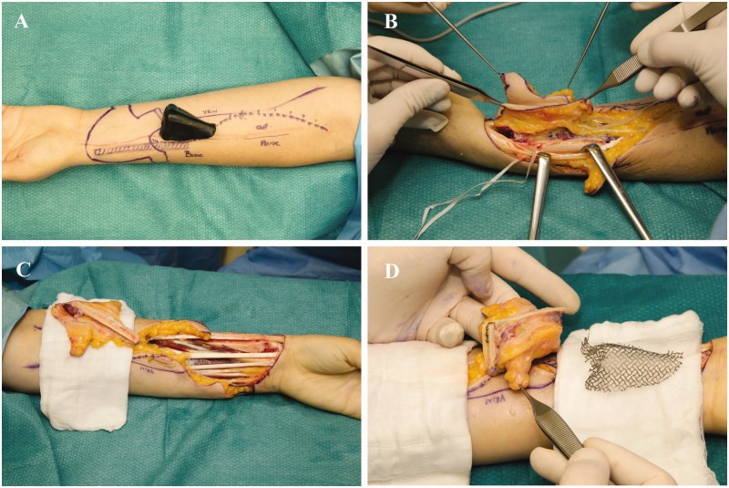
10.5.2 Choosing a flap for the maxilla
The maxilla is where these other flaps are used most. It is a different problem from the mandible.
- The mandible is a single load-bearing arch, so the main need is a long, strong strut of bone. The fibula fits this well.
- The maxilla is thin and hollow. It supports the eye, separates the mouth from the nose and sinus, holds the upper teeth and gives the cheek its projection.
- A maxillary defect may therefore need closure of the palate, support for the orbital floor, bone for teeth, or only a prosthesis (obturator). Often it needs more soft tissue than bone.
What is needed depends on how much of the maxilla is removed. Brown and Shaw (2010) classify the defect in two directions:
- Vertical extent (class I to VI): how high the resection goes, from the palate up to the orbit.
- Horizontal extent (a to d): how much of the palate and alveolus is lost. a = palate only, alveolus intact; b = half or less of the alveolus and palate, on one side; c = half or less but crossing the midline, or the front of the maxilla; d = more than half.
The higher the defect, the more the flap must support the orbit, and the more bone and soft tissue it needs.
| Vertical class | Defect | Usual reconstruction |
|---|---|---|
| I | Maxillectomy not causing an oronasal fistula | Local flap or none |
| II | Low maxillectomy, not involving the orbit | Obturator, or soft tissue or bone flap (fibula, iliac crest, scapula) |
| III | Involving the orbital floor, eye preserved | Bone flap to support the orbit: iliac crest with internal oblique, scapula tip, fibula |
| IV | With orbital exenteration | Large composite flap: iliac crest, scapula with latissimus |
| V, VI | Orbitomaxillary; nasomaxillary | Soft tissue flap; radial forearm with bone for the nasal defect |
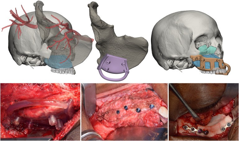
10.6 Vascularized bone for long bone defects
In the limbs, plastic surgeons provide vascularized bone for defects after tumor resection, trauma and infection.
The problem: a fibula is alive but thin compared with a femur or tibia. There are two solutions.
Flowchart: Fibula for a large long bone defect
- Fibula is alive but thin
- Add allograft around it
- Capanna techniqueallograft = strength, fibula = living bone
- Fold it
- Double-barrel fibulatwice the thickness
- Load it gradually
- Hypertrophy with time
- Add allograft around it
10.6.1 Fibula combined with allograft (Capanna technique)
In this technique a vascularized fibula is placed inside the canal of a large (massive) allograft. The two are used together to fill a long gap in the femur or tibia.
Where it came from
Massive allografts were widely used after bone tumor resection, but many failed through nonunion at the junctions, fracture or resorption. In Florence, Capanna and colleagues used a vascularized fibula to rescue these failed allografts, with very good results. From 1988 they combined the two grafts at the first operation, instead of waiting for the allograft to fail (Capanna 2007).
Why two grafts are better than one
| Massive allograft alone | Vascularized fibula alone | Both together | |
|---|---|---|---|
| Size and shape | Matches the femur or tibia | Thin compared with a femur or tibia | Allograft gives the size |
| Early strength | Good | Poor; fractures in about 1 of 3 adults (Elemosho 2025) | Allograft carries the load while the fibula grows |
| Biology | Dead; joins the host slowly or not at all | Living; unites like a fracture | Fibula unites at both junctions and revascularizes the allograft from inside |
| Over time | Fatigue fracture, infection, resorption | Hypertrophies under load | Fibula hypertrophies and fuses to the allograft |
In short, the allograft solves the mechanical problem and the fibula solves the biological problem.
Indications
- Intercalary defects of the femur or tibia longer than about 6 to 7 cm, mostly after resection of a bone sarcoma in a young patient who is expected to live long and be active (Houben 2019; Campanacci 2024).
- Resections next to a joint that leave less than about 3 cm of epiphysis, where the allograft gives a better hold for screws and for tendon reattachment (Campanacci 2024).
- Long post-traumatic defects. In 19 patients with distal femoral defects of 9.5 to 20 cm, 18 united at a mean of 6.6 months (Jayaramaraju 2019).
- Defects after chronic osteomyelitis when other methods have failed, as in the USM case below (Lam 2025).
- Congenital pseudarthrosis of the tibia in children (Van Den Heuvel 2020).
When it is usually not chosen: short defects (an allograft or fibula alone is enough), metastatic disease or short life expectancy, patients older than about 70 years, and some patients who will need radiotherapy after surgery (Campanacci 2024).
How it is done
- Plan. The resection length is measured on MRI. An allograft of matching size and side is ordered from the tissue bank, and the canal width is checked on CT so that the fibula will fit.
- Resect the tumor or dead bone. The orthopedic and plastic teams usually work at the same time: one resects while the other raises the fibula.
- Prepare the allograft. It is cut to length and the canal is reamed or burred out to take the fibula. A longitudinal slot is cut in the cortex so that the pedicle can leave the allograft without being kinked or compressed.
- Raise the fibula from the opposite leg, longer than the allograft. A skin paddle can be included for cover and monitoring.
- Assemble. The fibula is passed into the allograft. Its ends are pushed into the canal of the host bone at each end, so that living bone bridges both junctions.
- Fix. A long plate spans the allograft and both host segments. Screws are placed so that they avoid the fibula's pedicle. Long-spanning plates reduce later fractures (Campanacci 2024).
- Anastomose the peroneal vessels to local recipient vessels, then close the soft tissue or use the skin paddle.
If the canal of the allograft is too narrow for the fibula, for example in the distal tibia, the fibula is placed alongside the allograft (onlay) or combined with allograft cortical struts (Campanacci 2024).
Flowchart: How the combined graft heals
- Allograft carries the loadfrom the day of surgery; weight bearing is protected
- Fibula unites to the hostat both junctions, like a fracture; union at about 6 to 12 months
- Fibula hypertrophiesit thickens under load
- Fibula fuses to the allograftbone bridges form from the allograft's inner wall; the allograft is slowly revascularized from inside
This sequence was shown on serial X-rays and CT in 24 patients (Manfrini 2004). In 13 the allograft stayed intact and the fibula slowly enlarged and fused with it. In 8 the allograft fractured or failed to unite, and the fibula responded with rapid hypertrophy that healed the fracture. In 3 the fibula did not change in size or density and the reconstruction fractured without callus. This was taken to mean that the fibula had lost its blood supply. A fibula that does not hypertrophy is therefore a warning sign.
Variations
- Osteocutaneous fibula. Including a skin paddle covers the soft tissue lost at resection and allows the flap to be monitored. This was used in the USM series (Halim 2015).
- Chimeric fibula. The skin paddle is raised on a separate perforator so that it can be placed away from the bone (Lam 2025).
- "Capasquelet". An induced membrane is first created with a cement spacer (Masquelet). The allograft and fibula are then placed inside the membrane at the second stage. It has been reported in 4 patients with femoral defects, including infected cases (Combal 2021).
Evidence
| Study | Patients | Main results |
|---|---|---|
| Houben 2019, systematic review | 17 studies, 329 lower-limb reconstructions | Primary union 67 to 100%. Complications 6 to 86% and reoperation 10 to 92%, depending on the series. Good function (mean MSTS 24 to 29 of 30). |
| Othman 2020, meta-analysis: with fibula against allograft alone | 25 studies, lower-limb tumors | Adding the fibula lowered nonunion (13% vs 21%) and graft removal (7% vs 18%). Infection (8% vs 9%), fracture (20% vs 19%) and function were similar. |
| Elemosho 2025, meta-analysis: with allograft against fibula alone | 33 studies, 560 patients | In adults, adding the allograft lowered fibula fracture (17% vs 35%) but more patients needed reoperation (50% vs 27%). In children the two were similar. Chemotherapy raised the odds of nonunion about 10 times. |
| Houdek 2016, children | 18; mean age 11; mean follow-up 8 years | Limb salvage 94%. 14 needed another operation, 6 of them for nonunion. Mean MSTS 93%. 6 needed limb-length correction. |
| Campanacci 2024, tibia (Florence) | 74 fibula reconstructions, 55 with allograft; median follow-up 12 years | 49% had a complication needing surgery. Free from revision at 5 years: 58%. Reconstruction kept at 5 years: 91%. Nearly 10% ended in amputation. |
Interpretation. Adding a vascularized fibula to a massive allograft mainly reduces nonunion and the need to remove the graft. It does not prevent infection or fracture. Further operations are common in the first few years, but once the reconstruction has healed it lasts for decades. Patients and families should be told to expect a long course and probably more than one operation.
Complications
- Nonunion at one of the junctions, especially with chemotherapy. It is treated with bone graft and revision of fixation.
- Fracture of the allograft or of the plate, mostly in the first years.
- Infection of the allograft. Deep infection often means the allograft must be removed.
- Loss of the fibula's blood supply. The allograft then behaves like an allograft alone.
- Limb-length difference in growing children.
- Donor-site problems in the leg.
Aftercare
Weight bearing is protected until the junctions unite and is then increased step by step, because load is what makes the fibula hypertrophy. Union usually takes about 6 to 12 months; in the meta-analysis by Elemosho (2025), mean time to union was 9.4 months in children. X-rays are reviewed for union at both junctions and for thickening of the fibula.
USM experience
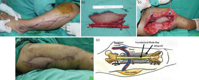
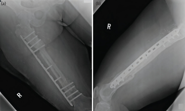
12 patients after bone tumor resection, reconstructed with free fibula osteocutaneous flap and massive allograft. Mean follow-up 63 months. Patients with lower limb reconstruction reached partial weight bearing at a mean of 4.2 months and full weight bearing at 8.2 months.
10.6.2 Double-barrel fibula
The fibula is folded into two parallel segments on one pedicle. This doubles the thickness.
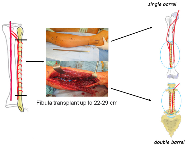
total talus reconstruction with a triangular double-barrel fibula after tumor resection in 4 patients. All achieved bony fusion. Median functional (MSTS) score was 74.5%.
10.6.3 Equipment and products used with bone flaps
| Purpose | Examples | Company |
|---|---|---|
| Planning software and printed guides | ProPlan CMF; IPS CaseDesigner | Materialise (with DePuy Synthes); KLS Martin |
| Reconstruction plates for the mandible | MatrixMANDIBLE; patient-specific plates | DePuy Synthes; KLS Martin; Stryker |
| Joining the vein without sutures | GEM Microvascular Anastomotic Coupler | Synovis (Baxter) |
| Monitoring a buried flap | Cook-Swartz implantable Doppler probe, placed around the vein or artery | Cook Medical |
| Monitoring through the skin paddle | Tissue oximeter (ViOptix T.Ox); handheld Doppler | ViOptix |
| Bone cuts with little heat | Piezoelectric saw (Piezosurgery) | Mectron |
10.7 Vascularized bone in the hand: scaphoid nonunion
The same principle applies on a small scale in the wrist.
The blood supply of the scaphoid enters distally and runs proximally. After a waist fracture, the proximal pole may lose its blood supply (avascular necrosis). A non-vascularized graft placed against dead bone often fails. Two vascularized grafts are used:
- Pedicled distal radius graft on the 1,2 intercompartmental supraretinacular artery (Zaidemberg 1991).
- Free medial femoral condyle graft on the descending genicular artery (Doi 2000).
Other pedicled vascularized grafts from the dorsal distal radius are used in the wrist, for example the 4,5 extensor compartmental artery graft for Kienböck disease of the lunate.
Jones (2008) compared them in 22 scaphoid nonunions with avascular proximal pole and carpal collapse. Union was achieved in 12 of 12 with the medial femoral condyle graft and 4 of 10 with the distal radius graft.
10.8 Vascularized epiphyseal transfer in children
The last special use is in the growing child. When a tumor resection removes the growth plate (for example at the distal radius or proximal humerus), an ordinary fibula flap will not grow.
In vascularized fibular epiphyseal transfer, the proximal fibula is taken with its growth plate and joint surface. It is based on the anterior tibial artery, because this vessel supplies the proximal fibular epiphysis.
- The commonest complications are peroneal nerve palsy and fracture of the flap (Lee 2023).
- Growth is expected but cannot be guaranteed. The family must be told this.
The technique was developed by Innocenti and colleagues in Florence. Their series grew over more than two decades, and the later papers give longer follow-up and larger numbers:
| Study | Patients | Main findings |
|---|---|---|
| Innocenti 1998 | 12 children under 10 years (9 proximal humerus, 3 distal radius) | First series. Grafts on the anterior tibial artery grew more than 1 cm a year (0.75 to 1.33 cm). Two other grafts were based on the peroneal artery. |
| Innocenti 2004 | 6 children, distal radius, mean age 8.4 years; mean follow-up 4.4 years | All united within 2 months. The 5 followed for 3 years or more grew steadily, and the new joint surface remodeled. Function excellent in all but 1. Peroneal nerve palsy in 3 (persistent in 1). |
| Innocenti 2005 (harvest technique) | 24 children under 11 years, upper limb | Detailed technique of harvesting the proximal fibula on the anterior tibial artery |
| Innocenti 2007 | 27 children aged 2 to 11 years; follow-up 2 to 14 years | Review of the whole Florence experience: indications and limits of the method |
| Innocenti 2015 (long-term) | 8 children, distal radius, mean age 7.3 years; 6 followed for a mean of 13.2 years (8 to 22) | All survived and united. Growth 0.7 to 1.4 cm a year; growth arrest in only 1, after trauma. After up to 22 years the wrist was painless, without degenerative change, with nearly normal motion. |
| Azoury 2023 (international, multicenter, with Innocenti) | 27 children, proximal humerus, median age 7 years; mean follow-up 10 years | 2 flap failures (7.4%). Growth in 24 of 27 (88.9%), mean 0.83 cm a year. Fracture of the transferred bone in 11 (40.7%), only 1 needing surgery. Peroneal nerve palsy in 16 (59.3%), permanent in 3 (11.1%). |
Interpretation. The later and larger series confirm the early result: a proximal fibula on the anterior tibial artery survives, unites, keeps growing at about 1 cm a year and gives a joint surface that lasts. They also show the price. Peroneal nerve palsy is common, because the nerve winds around the fibular neck next to the anterior tibial vessels, although most cases recover within a year. In the proximal humerus, fracture of the transferred fibula is common in the first years. The technique has since been used for the mandibular ramus and condyle in a 13-year-old after osteosarcoma resection, with an open growth plate at one year (Innocenti 2020).
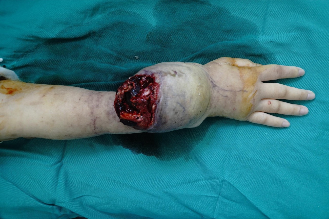
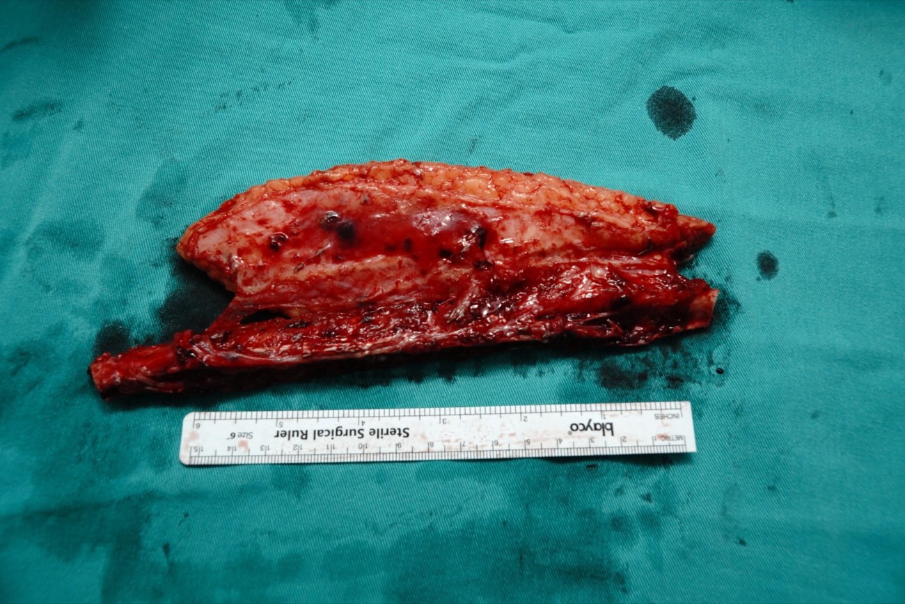
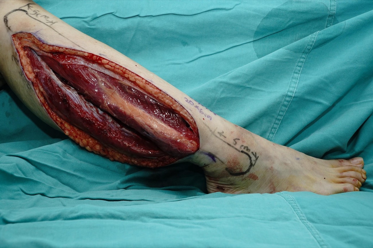
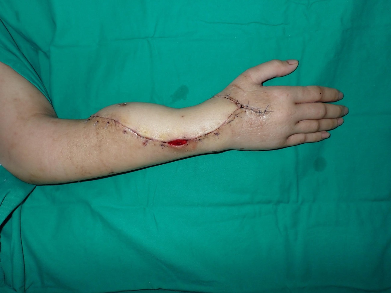
Figure 10.10. USM case: giant cell tumor of the distal radius. Tumor, resected specimen, fibula harvest and result.
10.9 USM papers on vascularized bone
The reconstructive team at Universiti Sains Malaysia (USM), Kubang Kerian, has published nine papers on vascularized bone. They cover the mandible, long bones, the foot, the skull and the donor leg.
| Paper | Site | Design | Patients |
|---|---|---|---|
| Chai 2019 | Mandible (ameloblastoma) | Retrospective series | 27 |
| Imran 2004 | Long bones: flap monitoring | Case series | 13 |
| Imran 2003 | Long bones: time to union | Case series | 13 |
| Halim 2015 | Long bones after tumor resection | Retrospective series | 12 |
| Lam 2025 | Femur (chronic osteomyelitis) | Case report | 1 |
| Rosli 2022a | Talus after tumor resection | Retrospective series | 4 |
| Sulaiman 2015 | Donor leg in children | Long-term follow-up | 8 |
| Rosli 2022b | Donor leg: peronea arteria magna | Case report | 1 |
| Ali 2005 | Orbit and frontal bone in a child | Case report | 1 |
10.9.1 Mandible: 20 years of fibula reconstruction after ameloblastoma
A 20-year experience of immediate mandibular reconstruction using free fibula osteocutaneous flaps following ameloblastoma resection: radical resection, outcomes, and recurrence.
Koh Siang Chai, Farah Hany Omar, Arman Zaharil Mat Saad, Wan Azman Wan Sulaiman, Ahmad Sukari Halim.
Archives of Plastic Surgery 2019;46(5):426–432.
What was done. All patients with ameloblastoma of the mandible treated from August 1997 to August 2017 were reviewed. Each had resection with a 2 cm margin (segmental mandibulectomy, or hemimandibulectomy for large tumors near the ramus). Each had immediate reconstruction with a free fibula osteocutaneous flap. The fibula was cut into two or three segments and fixed to a pre-bent plate at the lower border of the mandible. In later cases a 3D model was used to bend the plate.
What was found.
- 27 patients; mean age 29.7 years. Tumors were large, with a mean size of 6.4 × 4.9 cm, because patients presented late.
- A mean of 22.6 cm of fibula was harvested and 13.1 cm was used. The facial artery was the commonest recipient artery. Mean ischemia time was 169 minutes.
- All flaps survived. Two (7.4%) needed re-exploration for venous thrombosis. One had a recipient-site infection.
- No tumor recurred during a mean follow-up of 5.6 years.
- Only 2 patients had dental implants, because of cost. Most dentures were later abandoned because they did not fit the reconstructed jaw; patients chewed on the healthy side.
Interpretation. Wide resection followed by immediate fibula reconstruction is reliable in this population, which is younger than in Western series and presents with larger tumors. The paper reports survival of the flap and control of the tumor. It does not measure chewing, speech or bone height. The low use of dental implants shows that a fibula placed at the lower border is often not enough for dental rehabilitation; the bone may need to be placed higher or double-barreled. Ameloblastoma can recur late, so 5.6 years is not long enough to be sure of cure. The authors recommend yearly review for life.
10.9.2 The skin paddle as a monitor of buried bone
Skin paddle as an indicator of the viability of vascularised fibular graft.
Y Imran, Zulmi Wan, Ahmad Sukari Halim.
Singapore Medical Journal 2004;45(3):110–112.
What was done. 13 patients had long bone reconstruction with a free fibula that carried a skin paddle. The skin paddle was used to watch the flap after surgery, instead of bone scan or angiography.
What was found. All 4 anastomotic failures (100%) were seen early through changes in the skin paddle. Overall, 12 of 13 flaps (92%) succeeded, so 3 of the 4 failing flaps were saved.
Interpretation. A visible skin paddle is a simple, cheap and continuous monitor for bone that cannot be seen. The paddle shows the state of its own perforator. In most cases this reflects the pedicle, but a paddle can fail alone while the bone is still perfused. The study is small and has no comparison group. Its message agrees with larger series, in which buried flaps without a monitor were found late and could not be saved.
10.9.3 Time to union of a vascularized fibula in long bones
Skeletal union following long bone reconstruction using vascularised fibula graft.
Y Imran, Zulmi Wan, Ahmad Sukari Halim.
Singapore Medical Journal 2003;44(6):286–287.
What was done. 13 patients had a vascularized fibula after resection of diseased bone: 11 in the lower limb and 2 in the upper limb. Union was judged clinically and on X-ray.
What was found. Mean time to union was 32 weeks (earliest 8 weeks). Six of the 11 lower-limb patients (54%) had started weight bearing, at a mean of 27 weeks.
Interpretation. Living bone unites, but in a long bone it still takes months. The limb must be protected, and load is added slowly so that the fibula can hypertrophy. The two Imran reports appear to describe the same 13 patients. They are short reports with mixed diagnoses.
10.9.4 Fibula with massive allograft after bone tumor resection
Long-term outcome of free fibula osteocutaneous flap and massive allograft in the reconstruction of long bone defect.
Ahmad Sukari Halim, Siew Cheng Chai, Wan Faisham Wan Ismail, Wan Azman Wan Sulaiman, Arman Zaharil Mat Saad, Zulmi Wan.
Journal of Plastic, Reconstructive & Aesthetic Surgery 2015;68(12):1755–1762.
What was done. 12 consecutive patients with primary bone tumors had single-stage reconstruction with a massive allograft and a free fibula osteocutaneous flap. Unlike the original Capanna technique, the fibula carried a skin paddle. All were followed for at least 24 months.
What was found.
- Mean age 19.8 years. Eight had malignant tumors (five osteosarcoma, two chondrosarcoma, one synovial sarcoma) and four had benign tumors.
- Mean follow-up was 63 months (24 to 124 months).
- After lower-limb reconstruction, patients reached partial weight bearing at a mean of 4.2 months and full weight bearing at 8.2 months.
Interpretation. The allograft gives size and early strength; the fibula gives living bone that unites the allograft to the host. The skin paddle adds cover for the soft tissue lost at tumor resection and acts as a monitor. The abstract does not give rates of union, infection or allograft fracture, and there is no comparison group. The result shows what is possible in a small group of selected, young patients.
10.9.5 Chimeric fibula and allograft for a femur lost to osteomyelitis
Chimeric free fibula osteocutaneous flap and massive allograft for refractory post-traumatic osteomyelitis femur defect: a case report.
H Y Lam, Ahmad Sukari Halim, Wan Azman Wan Sulaiman.
Malaysian Orthopaedic Journal 2025;19(1):127–130.
What was done. A 31-year-old woman had infection after plating of a femoral shaft fracture. Implant removal, external fixation and an antibiotic cement spacer all failed. Final debridement left a 15 cm femoral defect and a scarred lateral thigh. A 15 cm fibula was placed inside a massive allograft and fixed with a locking plate. There was no septocutaneous perforator, so the skin paddle was raised on a separate muscle perforator (a chimeric flap). The bone could then be inset from the medial side, near the profunda femoris for the anastomosis, while the skin reached the lateral thigh.
What was found. A hematoma pressing on the perforator of the skin paddle was evacuated on the first day. The flap survived. At two years the femur had united and the infection had settled.
Interpretation. The Capanna principle can be used for infection when other options have failed. A chimeric design solves two problems: no septocutaneous perforator, which occurs in about 5 to 10% of legs, and skin needed in a different place from the bone. It is one case, and placing allograft in a previously infected bed carries a risk of late infection. Longer follow-up is needed.
10.9.6 Total talus reconstruction with a triangular double-barrel fibula
Functional outcomes in total talus reconstruction with triangular double-barrel free fibula flap following oncological resection: a retrospective case series review.
Mohamad Aizat Rosli, Wan Azman Wan Sulaiman, Wan Faisham Wan Ismail, Sahran Yahaya, Arman Zaharil Mat Saad, Zulmi Wan, Ahmad Sukari Halim.
Journal of Plastic, Reconstructive & Aesthetic Surgery 2022;75(2):641–650.
What was done. Patients who had the whole talus removed for tumor between 2009 and 2019 were reviewed. The fibula was cut into two struts and set as a wide-based triangle to replace the talus. Function was scored with the Musculoskeletal Tumour Society (MSTS) score.
What was found.
- Four patients; median age 32; median follow-up 60 months (24 to 132).
- Two flaps had venous thrombosis; both were saved at re-exploration.
- All four achieved solid bony fusion. Median MSTS score was 74.5% (66 to 76%).
- No donor-site problems and no local recurrence.
Interpretation. A living, fused hindfoot is a biological alternative to amputation after total talectomy. The triangle gives a broad, stable base. With four patients, the result is a proof that the technique works, not a measure of how often it works. Half the flaps needed re-exploration, so close monitoring is essential.
10.9.7 The donor ankle in children
Long-term effect on foot and ankle donor site following vascularized fibular graft resection in children.
Abdul Razak Sulaiman, Zulmi Wan, Shukrimi Awang, Aminudin Che Ahmad, Ahmad Sukari Halim, Rajaei Ahmad Mohd Zain.
Journal of Pediatric Orthopaedics B 2015;24(5):450–455.
What was done. Eight children aged 3 to 12 years had a fibula harvested, always leaving at least 6 cm of distal fibula. They were reviewed 3 to 12 years later.
What was found.
- Two children who had the middle shaft removed at 3 and 5 years of age developed ankle valgus from abnormal growth of the distal tibia. They needed growth modulation and a supramalleolar osteotomy with tibiofibular synostosis.
- One child who had the whole proximal fibula removed at 6 years developed hindfoot valgus from weak tibialis posterior. He needed talonavicular fusion and tendon transfer.
- Children older than 8 years at surgery had no deformity.
Interpretation. The rule of leaving 6 cm of distal fibula protects the adult ankle. It does not fully protect the ankle of a child under 6 years. Families must be told of this risk, and the child should be followed until growth ends. The study is small, but the deformities were serious and needed further surgery.
10.9.8 Harvesting a fibula in peronea arteria magna
A lucky case of successful free fibula osteocutaneous flap harvest in peronea arteria magna.
Mohamad Aizat Rosli, Wan Azman Wan Sulaiman, Ahmad Sukari Halim.
Archives of Plastic Surgery 2022;49(2):253–257.
What was done. A 17-year-old had a recurrent foot tumor resected, leaving an 8 × 8 × 4 cm defect. Pulses were normal, and planning used only a handheld Doppler. During harvest, the peroneal artery was found to be the main supply to the foot: the posterior tibial artery was aplastic and the anterior tibial artery hypoplastic (Kim-Lippert type III-C). Before dividing the vessel, the tourniquet was released and the peroneal artery was clamped for 15 minutes. The dorsalis pedis pulse stayed present and oxygen saturation of the foot stayed at 100%, so the flap was taken.
What was found. There was no ischemia of the donor foot. At six months the bone had united and the patient could bear full weight.
Interpretation. Normal pulses do not exclude peronea arteria magna, because collaterals from a large peroneal artery can fill both pulses. A clamp test before dividing the peroneal artery is a useful safety step when the anatomy looks unusual. The authors saw this variant once in 51 fibula flaps over five years. They do not image routinely, but they use CT angiography after leg trauma or with vascular disease. The title itself says the outcome was lucky; harvest in this anatomy remains a risk to the leg.
10.9.9 Vascularized calvarial bone for the orbit in a child
Combination of vascularized outer-table calvarial bone graft based on the superficial temporal vessels and allomatrix for the repair of an orbito-frontal blow-out fracture in a child.
Faizal Ali, Ahmad Sukari Halim, Sharida Zakaria Najihah, Mohtar Ibrahim, Jafri Abdullah.
Journal of Cranio-Maxillofacial Surgery 2005;33(5):326–330.
What was done. A 7-year-old girl had a traumatic orbito-frontal bone defect (Posnick type 2). It was repaired with outer-table calvarial bone kept on the superficial temporal vessels. The gaps were filled with Allomatrix, a calcium sulfate putty containing demineralized bone matrix.
What was found. Four years later the cosmetic result was good, with an artificial left eye.
Interpretation. A pedicled calvarial flap brings living bone to the orbit and forehead without microsurgery. It is most useful where the bed is scarred or irradiated, and it may grow with the child. It is one case, and growth was not measured.
10.10 Summary
- Vascularized bone stays alive, heals like a fracture and does not depend on the bed.
- It is used for long defects, poor beds, composite defects and avascular nonunion.
- The fibula is the main flap. It survives osteotomy because of its periosteal supply.
- It still needs bone contact, stable fixation and good soft tissue closure.
References
- Taylor GI, Miller GD, Ham FJ. The free vascularized bone graft: a clinical extension of microvascular techniques. Plast Reconstr Surg 1975;55:533–544.
- Wei FC, Chen HC, Chuang CC, Noordhoff MS. Fibular osteoseptocutaneous flap: anatomic study and clinical application. Plast Reconstr Surg 1986;78:191–200.
- Hidalgo DA. Fibula free flap: a new method of mandible reconstruction. Plast Reconstr Surg 1989;84:71–79.
- Chai KS, Omar FH, Mat Saad AZ, Wan Sulaiman WA, Halim AS. A 20-year experience of immediate mandibular reconstruction using free fibula osteocutaneous flaps following ameloblastoma resection. Arch Plast Surg 2019;46:426–432.
- de Boer HH, Wood MB. Bone changes in the vascularised fibular graft. J Bone Joint Surg Br 1989;71:374–378.
- Imran Y, Wan Z, Halim AS. Skin paddle as an indicator of the viability of vascularised fibular graft. Singapore Med J 2004;45:110–112.
- Brown JS, Barry C, Ho M, Shaw R. A new classification for mandibular defects after oncological resection. Lancet Oncol 2016;17:e23–e30.
- Jewer DD, Boyd JB, Manktelow RT, et al. Orofacial and mandibular reconstruction with the iliac crest free flap: a review of 60 cases and a new method of classification. Plast Reconstr Surg 1989;84:391–403.
- Cordeiro PG, Disa JJ, Hidalgo DA, Hu QY. Reconstruction of the mandible with osseous free flaps: a 10-year experience with 150 consecutive patients. Plast Reconstr Surg 1999;104:1314–1320.
- Taylor GI, Townsend P, Corlett R. Superiority of the deep circumflex iliac vessels as the supply for free groin flaps. Plast Reconstr Surg 1979;64:595–604.
- Swartz WM, Banis JC, Newton ED, et al. The osteocutaneous scapular flap for mandibular and maxillary reconstruction. Plast Reconstr Surg 1986;77:530–545.
- Soutar DS, Scheker LR, Tanner NS, McGregor IA. The radial forearm flap: a versatile method for intra-oral reconstruction. Br J Plast Surg 1983;36:1–8.
- Capanna R, Campanacci DA, Belot N, et al. A new reconstructive technique for intercalary defects of long bones: the association of massive allograft with vascularized fibular autograft. Orthop Clin North Am 2007;38:51–60.
- Halim AS, Chai SC, Wan Ismail WF, Wan Sulaiman WA, Mat Saad AZ, Wan Z. Long-term outcome of free fibula osteocutaneous flap and massive allograft in the reconstruction of long bone defect. J Plast Reconstr Aesthet Surg 2015;68:1755–1762.
- Imran Y, Wan Z, Halim AS. Skeletal union following long bone reconstruction using vascularised fibula graft. Singapore Med J 2003;44:286–287.
- Lam HY, Halim AS, Wan Sulaiman WA. Chimeric free fibula osteocutaneous flap and massive allograft for refractory post-traumatic osteomyelitis femur defect. Malays Orthop J 2025;19:127–130.
- Rosli MA, Wan Sulaiman WA, Wan Ismail WF, Yahaya S, Mat Saad AZ, Wan Z, Halim AS. Functional outcomes in total talus reconstruction with triangular double-barrel free fibula flap following oncological resection. J Plast Reconstr Aesthet Surg 2022;75:641–650. [Rosli 2022a]
- Bienger K, Stefan V, Dragu A, Bota O, et al. Free vascularized fibula transfer in single- or double-barrel technique. GMS Interdiscip Plast Reconstr Surg DGPW 2024;13:Doc07.
- Innocenti M, Ceruso M, Manfrini M, Angeloni R, Lauri G, Capanna R, Bufalini C. Free vascularized growth-plate transfer after bone tumor resection in children. J Reconstr Microsurg 1998;14:137–143.
- Innocenti M, Delcroix L, Manfrini M, Ceruso M, Capanna R. Vascularized proximal fibular epiphyseal transfer for distal radial reconstruction. J Bone Joint Surg Am 2004;86:1504–1511.
- Innocenti M, Delcroix L, Romano GF. Epiphyseal transplant: harvesting technique of the proximal fibula based on the anterior tibial artery. Microsurgery 2005;25:284–292.
- Innocenti M, Delcroix L, Romano GF, Capanna R. Vascularized epiphyseal transplant. Orthop Clin North Am 2007;38:95–101.
- Innocenti M, Baldrighi C, Menichini G. Long term results of epiphyseal transplant in distal radius reconstruction in children. Handchir Mikrochir Plast Chir 2015;47:83–89.
- Azoury SC, Shammas RL, Othman S, Sergesketter A, Brigman BE, Nguyen JC, Arkader A, Weber KL, Erdmann D, Levin LS, Kovach SJ, Innocenti M. Outcomes following free fibula physeal transfer for pediatric proximal humerus reconstruction: an international multi-institutional study. Plast Reconstr Surg 2023;151:805–813.
- Innocenti M, Mori F, Raffaini M, Lucattelli E, Innocenti A. Mandibular ramus and condyle reconstruction with vascularized proximal fibular epiphyseal transfer in the pediatric patient: a case report. Microsurgery 2020;40:818–822.
- Lee CJ, Tiourin E, Khoshab N, et al. Vascularized fibular epiphyseal transfer for pediatric limb salvage. Plast Reconstr Surg Glob Open 2023;11:e5354.
- Sulaiman AR, Wan Z, Awang S, Che Ahmad A, Halim AS, Ahmad Mohd Zain R. Long-term effect on foot and ankle donor site following vascularized fibular graft resection in children. J Pediatr Orthop B 2015;24:450–455.
- Ali F, Halim AS, Najihah SZ, Ibrahim M, Abdullah J. Combination of vascularized outer-table calvarial bone graft based on the superficial temporal vessels and allomatrix for the repair of an orbito-frontal blow-out fracture in a child. J Craniomaxillofac Surg 2005;33:326–330.
- Rosli MA, Wan Sulaiman WA, Halim AS. A lucky case of successful free fibula osteocutaneous flap harvest in peronea arteria magna. Arch Plast Surg 2022;49:253–257. [Rosli 2022b]
- Zaidemberg C, Siebert JW, Angrigiani C. A new vascularized bone graft for scaphoid nonunion. J Hand Surg Am 1991;16:474–478.
- Doi K, Oda T, Soo-Heong T, Nanda V. Free vascularized bone graft for nonunion of the scaphoid. J Hand Surg Am 2000;25:507–519.
- Jones DB Jr, Bürger H, Bishop AT, Shin AY. Treatment of scaphoid waist nonunions with an avascular proximal pole and carpal collapse. J Bone Joint Surg Am 2008;90:2616–2625.
- Brown JS, Shaw RJ. Reconstruction of the maxilla and midface: introducing a new classification. Lancet Oncol 2010;11:1001–1008.
- Chim H, Salgado CJ, Mardini S, Chen HC. Reconstruction of mandibular defects. Semin Plast Surg 2010;24:188–197.
- Urken ML, Buchbinder D, Weinberg H, et al. Functional evaluation following microvascular oromandibular reconstruction of the oral cancer patient. Laryngoscope 1991;101:935–950.
- Manfrini M, Vanel D, De Paolis M, Malaguti C, Innocenti M, Ceruso M, et al. Imaging of vascularized fibula autograft placed inside a massive allograft in reconstruction of lower limb bone tumors. AJR Am J Roentgenol 2004;182:963–970.
- Houben RH, Rots M, van den Heuvel SCM, Winters HAH. Combined massive allograft and intramedullary vascularized fibula as the primary reconstruction method for segmental bone loss in the lower extremity: a systematic review and meta-analysis. JBJS Rev 2019;7:e2.
- Othman S, Bricker JT, Azoury SC, Elfanagely O, Weber KL, Kovach SJ. Allograft alone vs. allograft with intramedullary vascularized fibular graft for lower extremity bone cancer: a systematic review and meta-analysis. J Plast Reconstr Aesthet Surg 2020;73:1221–1231.
- Elemosho A, Czerniecki S, Ramadan S, Farhan S, Mitchell KS, Souza JM. Comparative outcomes of allograft with vascularized fibula graft vs vascularized fibula graft alone for post-oncologic lower extremity salvage: systematic review and meta-analysis. J Plast Reconstr Aesthet Surg 2025;108:75–85.
- Houdek MT, Wagner ER, Stans AA, Shin AY, Bishop AT, Sim FH, et al. What is the outcome of allograft and intramedullary free fibula (Capanna technique) in pediatric and adolescent patients with bone tumors? Clin Orthop Relat Res 2016;474:660–668.
- Campanacci DA, Scanferla R, Marsico M, Scolari F, Scoccianti G, Beltrami G, et al. Intercalary resection of the tibia for primary bone tumors: are vascularized fibula autografts with or without allografts a durable reconstruction? Clin Orthop Relat Res 2024;482:960–975.
- Jayaramaraju D, Venkataramani H, Rajasekaran RB, Agraharam D, Sabapathy SR, Rajasekaran S. Modified Capanna's technique (vascularized free fibula combined with allograft) as a single-stage procedure in post-traumatic long-segment defects of the lower end of the femur: outcome analysis of a series of 19 patients with an average gap of 14 cm. Indian J Plast Surg 2019;52:296–303.
- Van Den Heuvel SCM, Winters HAH, Ultee KH, Zijlstra-Koenrades N, Sakkers RJB. Combined massive allograft and intramedullary vascularized fibula transfer: the Capanna technique for treatment of congenital pseudarthrosis of the tibia. Acta Orthop 2020;91:605–610.
- Combal A, Thuau F, Fouasson-Chailloux A, Arrigoni PP, Baud'huin M, Duteille F, Crenn V. Preliminary results of the "Capasquelet" technique for managing femoral bone defects: combining a Masquelet induced membrane and Capanna vascularized fibula with an allograft. J Pers Med 2021;11:774.