9.1 Introduction
Fresh autograft is the only graft material, apart from vascularized bone, that has living bone-forming cells. An autograft is bone moved from one site to another in the same patient. In this chapter the bone is moved without its blood supply.
A non-vascularized graft has no blood supply after transfer. Most of its cells die. It must be revascularized from the recipient bed. So the result depends on the quality of the bed.
These are the commonest bone grafts in plastic surgery: alveolar bone graft in cleft, orbital floor repair, nasal dorsum reconstruction and bone grafting of facial fractures.
Flowchart: Overview of autogenous bone graft
- Autogenous bone graft (non-vascularized)
- Cancellous
- Fast revascularizationfully replaced by new bone
- Used for bone healingalveolar cleft, nonunion
- Donoriliac crest, tibia, distal radius
- Used for bone healingalveolar cleft, nonunion
- Fast revascularizationfully replaced by new bone
- Cortical
- Creeping substitutionslow; weak at 2 to 6 months
- Used for shape and supportorbit, nasal dorsum, skull
- Donorcalvarium, rib
- Used for shape and supportorbit, nasal dorsum, skull
- Creeping substitutionslow; weak at 2 to 6 months
- Corticocancellous
- Both propertiesiliac crest block
- Cancellous
9.2 Types of autograft: cancellous and cortical
There are two basic types. They are used for different purposes.
| Cancellous graft | Cortical graft | |
|---|---|---|
| Structure | Porous, with marrow; large surface area | Dense, compact |
| Living cells | Many on the surface; some survive | Few; most die |
| Strength at the time of grafting | None | Strong |
| Revascularization | Fast (days to weeks) | Slow (months) |
| Final result | Completely replaced by new bone | Never completely replaced; a mixture of dead graft and new bone |
| Used when we need | Bone healing and filling: alveolar cleft, nonunion, contained defects | Shape and support: orbital floor, nasal dorsum, cranial defects, strut grafts |
A corticocancellous graft has both, for example a block from the iliac crest.
9.3 How a graft incorporates
Incorporation is the process by which the graft is joined to and replaced by the patient's living bone. The two types incorporate differently.
- Cancellous graft: vessels grow quickly into the spaces. Osteoblasts first lay new bone on top of the dead trabeculae. Later the dead trabeculae are removed. Formation comes first, so the graft gets stronger from the start.
- Cortical graft: vessels cannot enter dense bone. Osteoclasts must first cut tunnels into the graft. Then osteoblasts fill the tunnels. Resorption comes first, so the graft becomes weaker for several months before it recovers. This slow process is called creeping substitution.
Flowchart: Incorporation of cancellous graft
- Graft placedsurface cells survive by diffusion
- Vessels grow indays to weeks
- New bone formed on dead trabeculae
- Dead trabeculae resorbed
- Fully replaced
Flowchart: Incorporation of cortical graft (creeping substitution)
- Graft placedmost cells die
- Osteoclasts cut tunnelsgraft becomes weaker
- Vessels enter the tunnels
- Osteoblasts fill tunnels with new bone
- Mixture of new bone and dead graft
| Time after grafting | Cancellous graft | Cortical graft |
|---|---|---|
| First week | Surface cells survive by diffusion; inflammation | Most cells die; graft is as strong as normal bone |
| 2 weeks to 2 months | Vessels have grown through the graft; new bone is laid on the dead trabeculae; X-ray density increases | Osteoclasts start cutting tunnels from the surface and ends; graft starts to weaken |
| 2 to 6 months | Dead trabeculae are being removed; graft is gaining strength | Weakest period: the graft is porous and can lose up to about half its strength |
| 6 to 12 months | Remodeled to normal bone; graft fully replaced | New osteons fill the tunnels; strength returns |
| After 1 to 2 years | Normal bone | Strong, but still a mixture of new bone and dead graft |
(Enneking 1975; Burchardt 1983)
What this means in practice. A cortical graft is weakest at about 2 to 6 months after surgery. A structural cortical graft must be protected by fixation during this time.
9.4 Graft volume in the face: which bone keeps its shape?
In the face and skull, autografts are used in two main ways. Some fill a gap, such as an alveolar cleft or a small defect after trauma or tumor surgery. Others are placed on the surface of the bone to restore contour, for example on the orbital rim, the cheek or the nose. Large defects after cancer resection usually need vascularized bone instead. For a graft used for contour, the main problem is not nonunion. It is loss of volume with time.
Three things decide how much volume a graft keeps:
- Structure: cortical bone keeps more volume than cancellous bone.
- Position: an inlay graft keeps more volume than an onlay graft.
- Fixation and soft tissue cover: a graft that is fixed rigidly, under healthy soft tissue, keeps more volume.
The embryological origin of the bone (membranous or endochondral) was once thought to be a fourth factor. The next section explains why it is not.
9.4.1 Why calvarial bone is preferred for onlay grafts
Surgeons noticed that calvarial grafts on the face kept their volume better than grafts from the iliac crest or rib. Two explanations were proposed, one after the other.
| First explanation (1983): the origin of the bone | Later explanation (1998): the structure of the bone | |
|---|---|---|
| Idea | Calvarium is membranous bone. Iliac crest and rib are endochondral bone. Membranous bone was thought to behave differently because of its origin, for example by being revascularized earlier. | Calvarium is mostly cortical bone. Iliac crest is mostly cancellous bone. Cortical bone keeps its volume better, whatever its origin. |
| Study | Zins and Whitaker compared membranous grafts with endochondral grafts. Volume lost: in rabbits 20% and 65%; in monkeys 17% and 88%. Membranous grafts were also revascularized earlier (Kusiak 1985). | Ozaki and Buchman separated the two factors. They compared cortical membranous, cortical endochondral and cancellous endochondral grafts. The two cortical grafts lost the same volume. The cancellous graft lost the most. |
| Weakness | The membranous graft was also the cortical graft. The study could not tell which factor caused the difference. | Animal study; but it answered the question the first study could not |
Current understanding. What matters is whether the graft is cortical or cancellous, not whether it is membranous or endochondral (Ozaki and Buchman 1998). The calvarium is still the usual first choice for an onlay graft, but for practical reasons: it gives cortical bone of the right thickness, it is often in the same operative field, and the scar is hidden in the hair.
9.4.2 Inlay and onlay grafts
Grafts of the same type kept more volume as inlay grafts than as onlay grafts, at every time point (Sugg 2013).
| Inlay graft | Onlay graft | |
|---|---|---|
| Where it lies | Inside a defect, with host bone on several sides | On the surface of a bone |
| Contact with host bone | On several sides, so vessels and cells enter from several sides | On one side only |
| Load | Carries load as part of the skeleton, so it is maintained | Carries little load, so it is resorbed |
| Soft tissue | Little pressure on it | Pressed on by the overlying skin and muscle |
| Examples | Alveolar cleft, gap after an osteotomy | Augmentation of the cheek, chin, nasal dorsum or forehead |
9.4.3 Choosing the graft
Flowchart: Choosing the type of graft in the face
- What is the graft for?
- To add contour (onlay)
- Volume must be kept
- Cortical graftcalvarium, outer table; fixed with a screw
- Volume must be kept
- To fill a gap (inlay)
- Bone must form quickly
- Cancellous graftiliac crest; packed firmly, covered with healthy soft tissue
- Bone must form quickly
- To support a thin wall
- Shape and stiffness
- Thin cortical graftsplit calvarium or rib, for the orbital floor or nasal dorsum
- Shape and stiffness
- To add contour (onlay)
Practical rules for onlay grafts in the face. Use cortical bone. Fix it rigidly, usually with a lag screw. Place it in contact with a bleeding bone surface, under well-vascularized soft tissue. Expect some loss of volume, more where the overlying soft tissue is tight or mobile (Oppenheimer 2008).
9.5 Requirements of the recipient bed
Because a non-vascularized graft depends on the bed, the bed must be prepared properly:
- Blood supply: bleeding bone surface under the graft, and well-vascularized soft tissue over it.
- Contact: wide contact between graft and host bone, with no dead space.
- Rigid fixation: Phillips and Rahn (1988) placed onlay grafts on sheep mandibles, with and without lag screw fixation. Fixed grafts kept more volume (86% for fixed skull grafts) and revascularized faster. A graft that moves resorbs.
- Soft tissue cover: closed without tension. An exposed graft is usually lost.
- No infection, no radiation. In an irradiated bed, use vascularized bone.
Handling: keep the graft moist (in blood or saline-soaked gauze) and place it without delay. Do not let it dry.
9.6 Donor sites
The donor site is chosen according to the type and amount of bone needed.
| Donor site | Type of bone | Main uses | Main risks |
|---|---|---|---|
| Calvarium (parietal) | Cortical (membranous) | Orbit, nasal dorsum, zygoma, forehead, cranial defects | Inner table breach, dural tear, bleeding |
| Iliac crest (anterior; posterior for more volume) | Cancellous and corticocancellous | Alveolar cleft, nonunion, mandibular defects, interpositional grafts | Pain, hematoma, lateral femoral cutaneous nerve injury, hernia |
| Rib | Cortical; with cartilage as costochondral graft | Skull defects in children; mandibular condyle and ramus | Pleural tear; unpredictable growth of costochondral graft |
| Proximal tibia | Cancellous | Alveolar and sinus grafting; small nonunions | Fracture (rare); avoid the growth plate in children |
| Distal radius, olecranon | Cancellous, small volume | Hand: scaphoid and phalangeal nonunion | Small volume |
| Mandibular symphysis and ramus | Cortical and corticocancellous, small | Alveolar ridge, small facial defects | Mental nerve and tooth root injury |
(Tessier 1982; Younger and Chapman 1989)
9.6.1 Calvarial bone graft
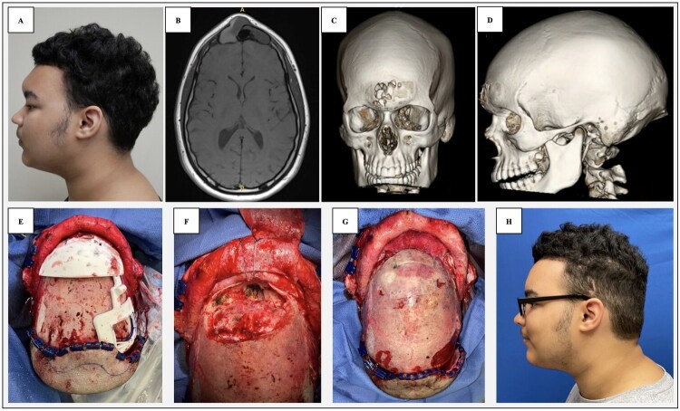
Calvarial bone is the first choice for the face: it is cortical, it is in the same operative field, and the scar is hidden in the hair. The skull has three layers: outer table, diploë and inner table. The graft is taken by splitting through the diploë.
Figure 9.3. Split calvarial graft.
- Site: parietal bone, behind the coronal suture and away from the midline (to avoid the sagittal sinus). The parietal bone is the thickest.
- In situ split: the outer table is lifted off, leaving the inner table. Possible in adults, who have a diploë.
- Ex vivo split: a full-thickness piece is removed, split on the table, and the inner table is put back. Used in young children, who do not yet have a diploë.
- Risks: breach of the inner table, dural tear, bleeding. Tessier and colleagues (2005) described the technique and complications from a combined experience of 9,650 cases.
Anatomy for safe harvest: the adult parietal bone is on average about 6 to 7 mm thick, but it varies widely, and CT is useful before harvest. The bone is thin in the temporal region and at the sutures. Harvest stays at least 1.5 to 2 cm from the midline to avoid the sagittal sinus and its lateral lakes. A reliable diploë is usually present from later childhood.
9.6.2 Iliac crest graft
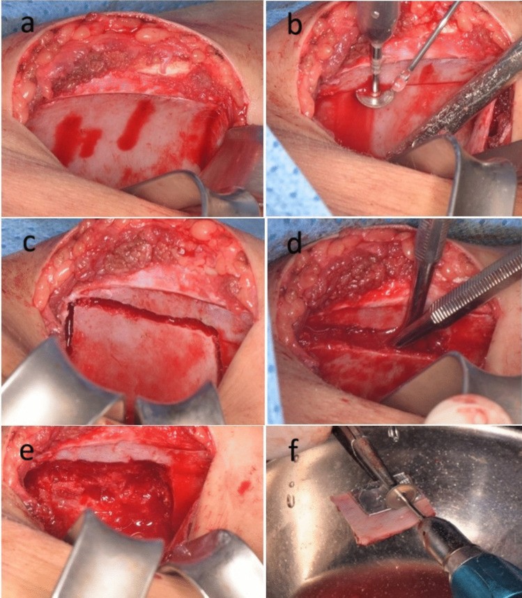
The iliac crest is the main source of cancellous bone. Complications are donor site pain, hematoma, injury to the lateral femoral cutaneous nerve and contour defect. In children the cartilage cap of the crest is preserved.
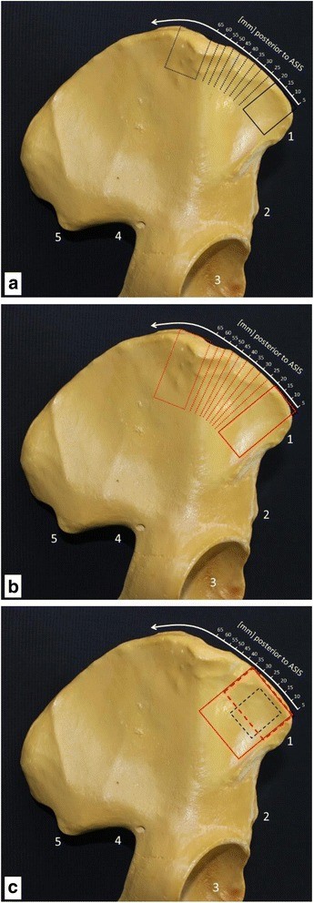
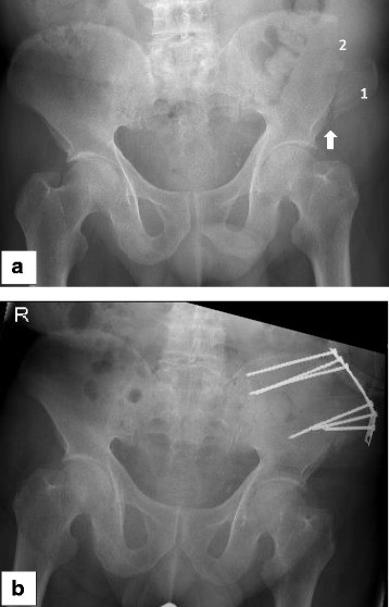
- Anterior crest: incision placed below or behind the crest, starting at least 2 to 3 cm behind the anterior superior iliac spine. This protects the lateral femoral cutaneous nerve and the inguinal ligament and sartorius attachments, and avoids avulsion fracture of the spine. Cancellous bone is taken through a cortical window or "trapdoor", or by trephine.
- Posterior crest: gives about twice the volume of the anterior crest. The patient must be prone. At risk: superior cluneal nerves, sciatic notch and superior gluteal artery, sacroiliac joint.
- Complications: in 243 grafts, major complications occurred in 8.6% and minor in 20.6% (Younger and Chapman 1989). The commonest long-term problem is donor site pain. Others: hematoma, seroma, infection, meralgia paresthetica, gait disturbance, fracture of the ilium, hernia through a full-thickness defect, contour deformity.
9.6.3 Rib and costochondral graft
- Harvested subperiosteally, usually the 5th to 7th rib, through an inframammary incision. Check for a pleural tear with a Valsalva maneuver under saline.
- Split rib is used for skull defects in children. The rib regenerates if the periosteum is kept.
- Costochondral graft: rib with a 2 to 3 mm cap of cartilage, used to reconstruct the mandibular condyle and ramus in children (hemifacial microsomia, TMJ ankylosis). It has growth potential, but growth is unpredictable, and overgrowth can occur.
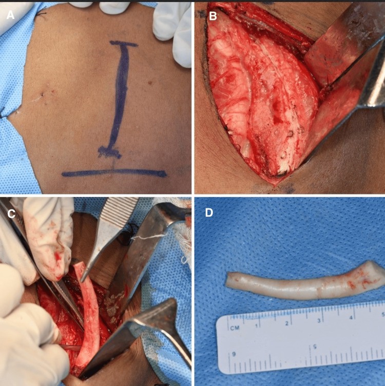
9.6.4 Instruments used to harvest and prepare the graft
The instrument decides the form of the graft: block, chips or fine particles. It also affects how many cells survive. Heat from a fast burr kills osteocytes, so sharp hand instruments, low-speed drills with irrigation and piezoelectric saws are preferred.
| Instrument | What it does | Typical use |
|---|---|---|
| Osteotome, gouge and curette | Lift a cortical window or block, then scoop cancellous bone | Iliac crest, calvarium, tibia |
| Trephine | A hollow drill that removes a core of cancellous bone through a small incision | Iliac crest for alveolar bone graft; chin; tibia |
| Piezoelectric saw | Cuts bone by ultrasonic vibration. It does not cut soft tissue, so nerves, dura and sinus lining are safer. | Block grafts from the ramus and chin; calvarial and orbital osteotomies |
| Bone scraper | A blade drawn over the cortex collects thin shavings in a chamber | Small volumes of particulate graft in the face and mouth |
| Bone mill | Grinds blocks or spare bone into particles of even size | Turning cortical or corticocancellous bone into packable graft; also used for resected bone that would be discarded |
| Reamer-irrigator-aspirator (RIA) | A reamer passed down the medullary canal of the femur or tibia, with continuous irrigation and suction. The reamings are caught in a filter. | Large volumes of cancellous-type graft for long bone defects, including the second stage of the induced membrane technique |
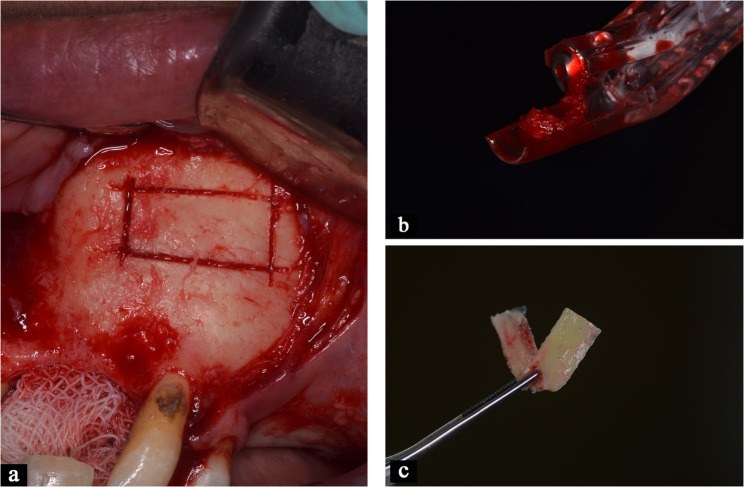
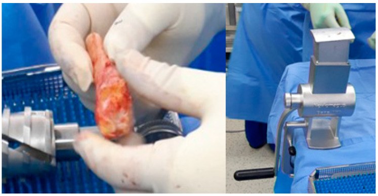
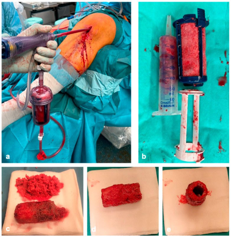
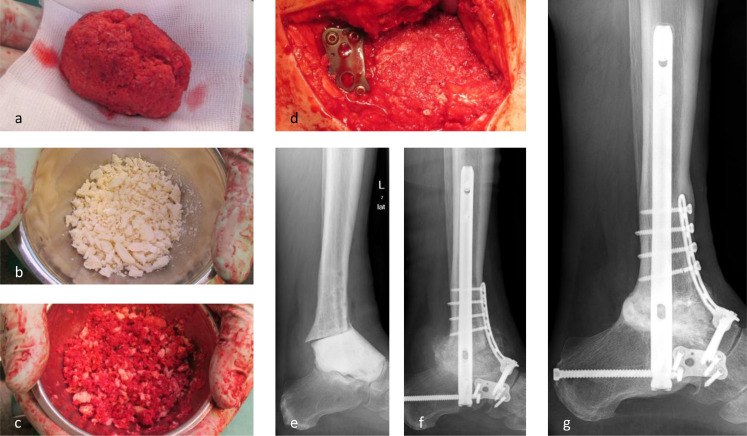
Examples of products used with autograft.
| Purpose | Product | Company |
|---|---|---|
| Harvest from the medullary canal | Reamer-Irrigator-Aspirator (RIA) | DePuy Synthes |
| Bone scraper for thin cortical shavings | SafeScraper | Meta |
| Piezoelectric bone saw | Piezosurgery | Mectron |
| Holding particulate graft together | Fibrin sealant, for example Tisseel | Baxter |
| Stopping bleeding from the cut bone | Bone wax; Ostene (a water-soluble substitute that does not block bone healing) | Several; Baxter |
| Fixing an onlay graft in a child | Resorbable plates and screws: LactoSorb, RapidSorb, SonicWeld Rx | Zimmer Biomet; DePuy Synthes; KLS Martin |
9.7 Alveolar bone graft
The alveolar bone graft is the commonest use of iliac cancellous graft in our specialty, so it is described here in detail.
Aims: to give a continuous dental arch, to give bone for the canine to erupt into, to close the oronasal fistula, to support the alar base, and to give bone for a dental implant if needed.
Why cancellous bone: the graft must be completely replaced by normal bone so that a tooth can erupt through it and be moved orthodontically. Cortical bone would not allow this.
Timing is classified by age:
| Type | Age | Comment |
|---|---|---|
| Primary | Under 2 years | Mostly abandoned: it restricts maxillary growth. |
| Early secondary | 2 to 5 years | Used in some centers. |
| Secondary | 9 to 11 years (mixed dentition) | Standard. Done before the canine erupts, when its root is about half to two-thirds formed. |
| Late secondary | After canine eruption | Poorer results. |
Evidence: Bergland (1986) reported 378 patients. The best results were when the graft was done at 9 to 11 years, before canine eruption: 64% achieved normal bone height between the teeth. The erupting tooth loads the graft, and loaded bone is maintained.
9.7.1 Technique
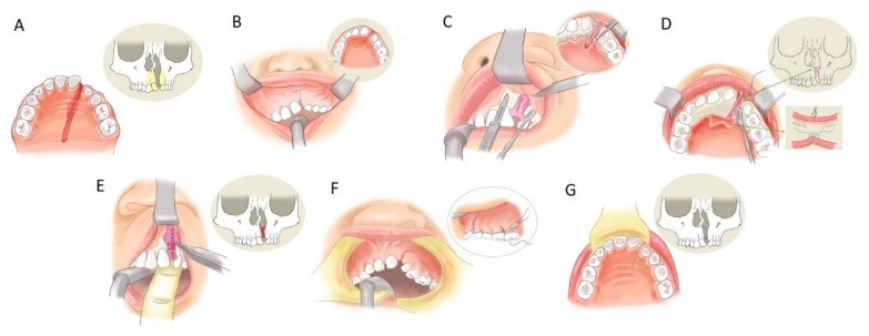
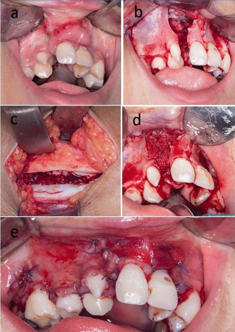
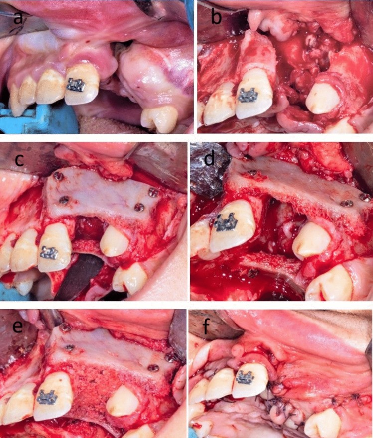
Flowchart: Steps of secondary alveolar bone grafting
- Orthodontic expansion of the arch
- Gingival mucoperiosteal flaps raised
- Nasal floor and palatal lining closedwatertight
- Cancellous iliac bone packed
- Flaps advanced and closed without tension
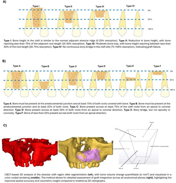
9.7.2 Assessment of the result
| Bergland type | Height of the bone between the teeth |
|---|---|
| I | Normal height |
| II | At least three-quarters of normal |
| III | Less than three-quarters of normal |
| IV | Failure: no continuous bony bridge |
Types I and II are counted as success. CT (cone beam) measures the volume more accurately than the plain radiographs used in the Bergland scale.
9.7.3 Factors that affect success
- Timing before canine eruption (the most important factor).
- Watertight closure of the nasal lining and tension-free gingival flap cover.
- Good oral hygiene and no active infection.
- Width of the cleft; bilateral clefts do worse than unilateral.
9.7.4 Alternatives to iliac crest
Tibia, mandibular symphysis and calvarium have been used; they give less cancellous bone. rhBMP-2 has been studied to avoid a donor site, mainly in skeletally mature patients, and is off-label.
9.8 Limits of non-vascularized graft: the mandible
The sections above show where non-vascularized graft works well. This section shows where it starts to fail. The best evidence is from segmental defects of the mandible.
| Study | Non-vascularized graft | Vascularized bone flap |
|---|---|---|
| Pogrel 1997 (68 patients) | 76% success (22 of 29) | 95% success (37 of 39) |
| Foster 1999 (75 patients) | 69% bony union (18 of 26) | 96% bony union (47 of 49) |
- In Pogrel's study, failure of non-vascularized grafts increased with the length of the defect. This is the source of the commonly quoted limit of about 6 cm.
- In Foster's study the vascularized group did better even though it had more irradiated patients (45% versus 11%).
Flowchart: Segmental mandibular defect: graft or flap?
- Segmental mandibular defect
- Short (under about 6 cm), lateral, clean bed, no radiotherapy
- Non-vascularized graft possibleunion about 70 to 75%
- Long, anterior, irradiated, contaminated, or implants planned
- Vascularized bone flapunion about 95%
- Short (under about 6 cm), lateral, clean bed, no radiotherapy
Summary. A non-vascularized graft is acceptable for a short lateral mandibular defect in a clean, non-irradiated bed. For a long defect, an anterior defect, an irradiated or contaminated bed, or when dental implants are planned, vascularized bone is needed.
References
- Enneking WF, Burchardt H, Puhl JJ, Piotrowski G. Physical and biological aspects of repair in dog cortical-bone transplants. J Bone Joint Surg Am 1975;57:237–252.
- Burchardt H. The biology of bone graft repair. Clin Orthop Relat Res 1983;174:28–42.
- Zins JE, Whitaker LA. Membranous versus endochondral bone: implications for craniofacial reconstruction. Plast Reconstr Surg 1983;72:778–785.
- Ozaki W, Buchman SR. Volume maintenance of onlay bone grafts in the craniofacial skeleton: micro-architecture versus embryologic origin. Plast Reconstr Surg 1998;102:291–299.
- Phillips JH, Rahn BA. Fixation effects on membranous and endochondral onlay bone-graft resorption. Plast Reconstr Surg 1988;82:872–877.
- Phillips JH, Rahn BA. Fixation effects on membranous and endochondral onlay bone graft revascularization and bone deposition. Plast Reconstr Surg 1990;85:891–897.
- Tessier P. Autogenous bone grafts taken from the calvarium for facial and cranial applications. Clin Plast Surg 1982;9:531–538.
- Tessier P, Kawamoto H, Posnick J, Raulo Y, Tulasne JF, Wolfe SA. Taking calvarial grafts, either split in situ or splitting of the parietal bone flap ex vivo: tools and techniques. V. A 9650-case experience in craniofacial and maxillofacial surgery. Plast Reconstr Surg 2005;116(5 Suppl):54S–71S.
- Younger EM, Chapman MW. Morbidity at bone graft donor sites. J Orthop Trauma 1989;3:192–195.
- Boyne PJ, Sands NR. Secondary bone grafting of residual alveolar and palatal clefts. J Oral Surg 1972;30:87–92.
- Bergland O, Semb G, Åbyholm FE. Elimination of the residual alveolar cleft by secondary bone grafting and subsequent orthodontic treatment. Cleft Palate J 1986;23:175–205.
- Pogrel MA, Podlesh S, Anthony JP, Alexander J. A comparison of vascularized and nonvascularized bone grafts for reconstruction of mandibular continuity defects. J Oral Maxillofac Surg 1997;55:1200–1206.
- Foster RD, Anthony JP, Sharma A, Pogrel MA. Vascularized bone flaps versus nonvascularized bone grafts for mandibular reconstruction. Head Neck 1999;21:66–71.
- Arrington ED, Smith WJ, Chambers HG, Bucknell AL, Davino NA. Complications of iliac crest bone graft harvesting. Clin Orthop Relat Res 1996;329:300–309.
- Buchman SR, Ozaki W. The ultrastructure and resorptive pattern of cancellous onlay bone grafts in the craniofacial skeleton. Ann Plast Surg 1999;43:49–56.
- Kusiak JF, Zins JE, Whitaker LA. The early revascularization of membranous bone. Plast Reconstr Surg 1985;76:510–516.
- Sugg KB, Rosenthal AH, Ozaki W, Buchman SR. Quantitative comparison of volume maintenance between inlay and onlay bone grafts in the craniofacial skeleton. Plast Reconstr Surg 2013;131:1014–1021.
- Oppenheimer AJ, Tong L, Buchman SR. Craniofacial bone grafting: Wolff's law revisited. Craniomaxillofac Trauma Reconstr 2008;1:49–61.