3.1 Introduction
Bone tissue has two parts: the matrix, which is the material between the cells, and the cells that make, maintain and remove it.
Every bone graft and every bone substitute is designed to supply one or more of these parts. A scaffold copies the matrix. Bone marrow aspirate supplies cells. BMP copies a signal stored in the matrix. Understanding the normal matrix and cells explains what each product can and cannot do.
Flowchart: Overview of bone tissue
- Bone tissue
- Matrix
- Mineralhydroxyapatite; stiffness
- Organic parttype I collagen and other proteins; toughness
- Water
- Cells
- Bone-forming linestem cell → osteoblast → osteocyte or lining cell
- Bone-removing lineblood-forming stem cell → osteoclast
- Matrix
3.2 The matrix: a composite material
Bone is a composite, like reinforced concrete. Each part does a different job:
| Part | Approximate share of weight | Main job |
|---|---|---|
| Mineral (hydroxyapatite) | About 60% | Stiffness; resists compression |
| Organic matrix (mainly type I collagen) | About 30% | Toughness; resists tension; stops cracks from spreading |
| Water | About 10% | Carries nutrients; lets the bone absorb energy |
The exact figures vary with age, site and how they are measured. Without mineral, bone bends like rubber. Without collagen, it shatters like chalk.
3.2.1 Mineral
The mineral is a form of calcium phosphate called hydroxyapatite, Ca10(PO4)6(OH)2. In bone the crystals are very small (nanometers) and imperfect: carbonate and other ions replace part of the phosphate. Small, imperfect crystals dissolve easily, which lets osteoclasts remove them and lets bone release calcium into the blood. Synthetic hydroxyapatite used as a bone substitute is more crystalline, and for that reason it dissolves very slowly.
3.2.2 Collagen and osteoid
About 90% of the organic matrix is type I collagen. Each molecule is a triple helix of two α1 chains and one α2 chain, coded by the genes COL1A1 and COL1A2. The molecules line up into fibrils and are joined by cross-links. Mineral crystals are laid down in the gaps within and between the fibrils.
Newly laid matrix that has not yet been mineralized is called osteoid. A thin seam of osteoid on a bone surface is normal: it shows that bone is being formed there.
In most types of osteogenesis imperfecta, type I collagen is abnormal. The bones contain mineral but break easily, which shows that the quality of the matrix matters as much as the amount of mineral.
3.2.3 Non-collagen proteins
The remaining 10% of the organic matrix is a mix of proteins. Some help mineral to form, some help cells to attach, and some are stored signals.
| Protein | Main role |
|---|---|
| Osteocalcin | Made by osteoblasts; binds mineral. Its blood level is a marker of bone formation. |
| Osteopontin, bone sialoprotein | Help cells attach to the matrix (osteoclasts attach through osteopontin); help mineral to start forming |
| Osteonectin | Links collagen and mineral |
| Proteoglycans | Organize collagen fibrils; bind growth factors |
| Growth factors: BMPs, TGF-β, IGF | Stored in the matrix and released when osteoclasts remove bone. This is why demineralized bone matrix (DBM) can induce bone: the mineral is removed and the stored BMPs are exposed. |
3.3 Mineralization
Laying down the matrix and mineralizing it are two separate steps.
Flowchart: How osteoid becomes bone
- Osteoblast lays down osteoidcollagen and other proteins
- Matrix vesicles bud off the osteoblastthe first crystals form inside them
- TNAP removes pyrophosphatepyrophosphate blocks crystal growth
- Primary mineralizationfast; most mineral within days to weeks
- Secondary mineralizationslow; continues for months
TNAP (tissue-nonspecific alkaline phosphatase) is the enzyme that splits pyrophosphate, a natural inhibitor of mineral growth. In hypophosphatasia, TNAP is defective, pyrophosphate builds up, and bone does not mineralize. The blood alkaline phosphatase is low. Replacing the enzyme (asfotase alfa) heals the bone disease (Whyte 2012).
Two pairs of terms are often confused:
- Ossification is the formation of bone tissue. Mineralization (calcification) is the deposit of mineral in a matrix. Calcified cartilage is mineralized but is not bone.
- Osteomalacia is a failure to mineralize osteoid: there is plenty of matrix but too little mineral. Osteoporosis is too little bone, with a damaged structure, but the bone that remains is normally mineralized.
3.4 The cells of bone
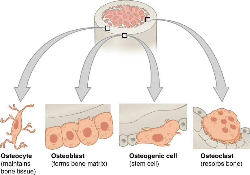
| Cell | Origin | Where it is | Function |
|---|---|---|---|
| Skeletal stem and progenitor cells | Mesenchyme | Periosteum (cambium layer), endosteum, marrow, cranial sutures | Supply new osteoblasts (and cartilage cells) for growth and repair |
| Osteoblast | Skeletal progenitor | On surfaces where bone is forming | Lays down osteoid and mineralizes it; controls osteoclasts |
| Lining cell | Resting osteoblast | Flat cells covering inactive surfaces | Covers the bone; can be reactivated |
| Osteocyte | Osteoblast buried in matrix | In lacunae | Senses load and damage; directs remodeling; controls phosphate |
| Osteoclast | Blood-forming stem cell (monocyte and macrophage line) | On surfaces where bone is being removed | Dissolves mineral and digests matrix |
3.4.1 Skeletal stem cells
New osteoblasts come from progenitor cells in the periosteum, the marrow and, in the skull, the sutures (Debnath 2018; Zhao 2015). These populations are not identical. Periosteal cells are especially important in fracture repair.
A "mesenchymal stem cell" (MSC) grown in a laboratory dish from marrow or fat is not the same as a true skeletal stem cell in the body. A true stem cell must renew itself and produce defined cell types. This difference matters when judging cell products sold for bone repair: a product may contain cells, but that does not prove that they will form bone.
3.5 The osteoblast
Two transcription factors (proteins that switch genes on) are essential for a progenitor to become an osteoblast:
- RUNX2. Mice without it form a cartilage skeleton with no bone at all (Komori 1997).
- Osterix (SP7), which acts after RUNX2 (Nakashima 2002).
The Wnt and BMP signaling pathways drive cells along this path.
Loss of one copy of RUNX2 causes cleidocranial dysplasia (Mundlos 1997): absent or small clavicles, open fontanelles and wide sutures, and many extra teeth that do not erupt. The bones affected are mostly membranous bones, which form directly from osteoblasts.
A mature osteoblast is a protein factory. It has a large rough endoplasmic reticulum and Golgi apparatus. It:
- makes collagen and the non-collagen proteins;
- releases matrix vesicles and alkaline phosphatase to start mineralization;
- makes RANKL and OPG, which control the osteoclast.
At the end of its working life (a few months), an osteoblast either is buried and becomes an osteocyte, flattens into a lining cell, or dies by apoptosis.
3.6 The osteocyte
The osteocyte was once thought to be a resting cell. It is now known to be the main controller of bone (Bonewald 2011). It does four things:
| Function | How |
|---|---|
| Senses load | Detects fluid flow in the canaliculi when bone bends |
| Brakes bone formation | Makes sclerostin, which blocks the Wnt pathway in osteoblasts. Loading reduces sclerostin, so bone forms where it is loaded. |
| Starts bone removal | Is the main source of RANKL in adult bone (Xiong 2011; Nakashima T 2011). A dying osteocyte near a microcrack signals osteoclasts to remove the damaged bone. |
| Controls phosphate | Makes FGF23, which makes the kidney excrete phosphate |
Osteocytes need blood supply. They die when bone loses its vessels: after a fracture, near a screw hole drilled with heat, or in a free bone graft. Dead bone keeps its shape, but it cannot sense load, repair itself or signal for its own replacement.
3.7 The osteoclast
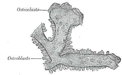
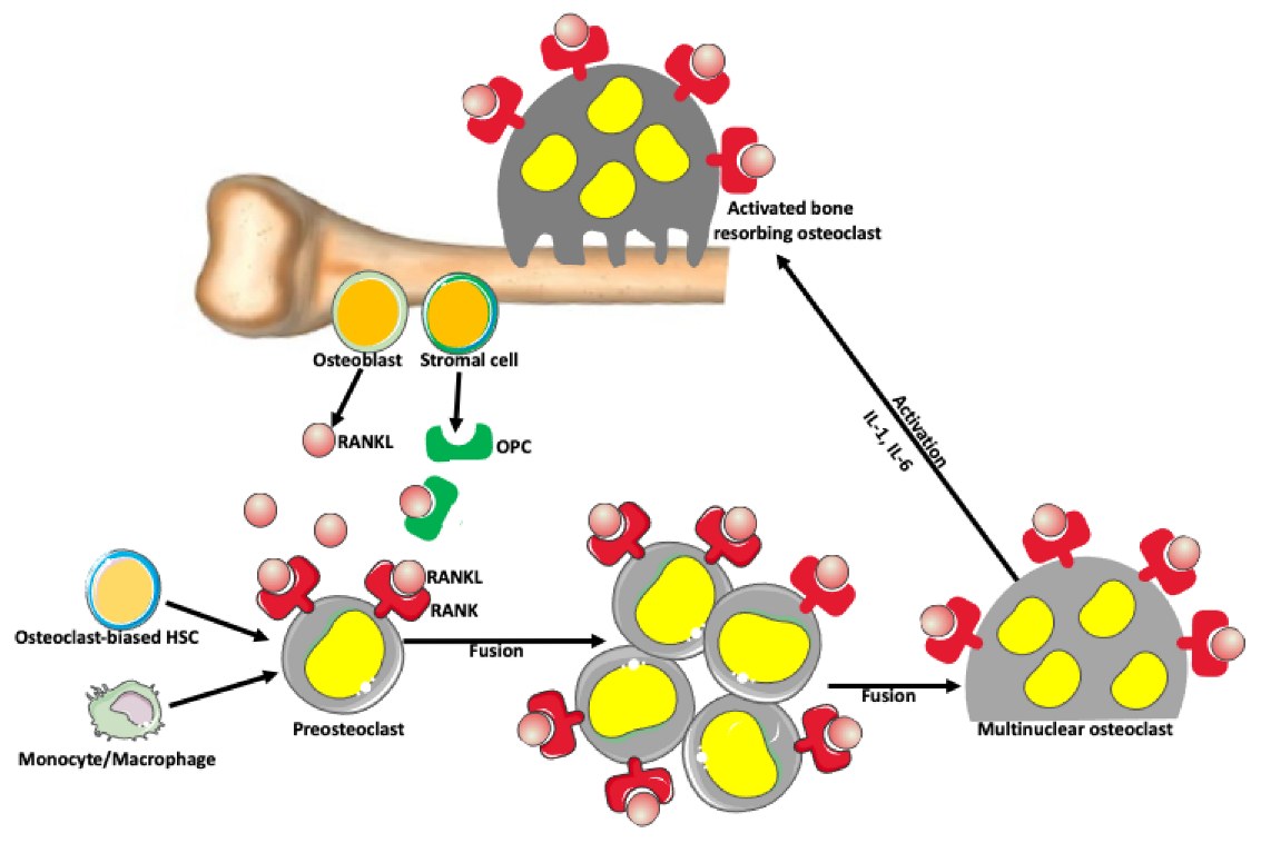
The osteoclast is a large cell with many nuclei (often 10 to 20), formed when precursor cells from the blood fuse together.
3.7.1 How an osteoclast is formed: RANKL, RANK and OPG
The osteoblast line controls the osteoclast through three molecules (Simonet 1997; Lacey 1998; Boyle 2003):
Flowchart: The RANKL, RANK and OPG system
- M-CSF keeps precursors alive
- Osteoblasts and osteocytes release RANKL
- RANKL binds RANKon the precursor cell
- Precursors fuse into osteoclasts
- Bone is removed
OPG (osteoprotegerin), also made by the osteoblast line, binds RANKL and stops it actingmore RANKL than OPG: more removal · more OPG than RANKL: less removal
Many things that cause bone loss act by raising RANKL: parathyroid hormone, lack of estrogen, glucocorticoids and inflammation. The drug denosumab is an antibody that blocks RANKL, in the same way as OPG.
3.7.2 How an osteoclast removes bone
- It attaches to the bone with a ring of actin called the sealing zone. This closes off a small space under the cell.
- The membrane facing the bone folds into a ruffled border, which greatly increases its surface.
- It pumps acid (hydrogen ions) into the sealed space through a proton pump. The acid dissolves the mineral.
- It releases enzymes, mainly cathepsin K, which digest the exposed collagen.
- It takes up the broken-down products and moves on, or dies.
The pit left behind is called a Howship lacuna. If osteoclasts fail, bone becomes very dense but brittle (osteopetrosis), and the marrow space is reduced. Patients with osteopetrosis are prone to osteomyelitis of the jaw, because their dense bone has a poor blood supply.
3.8 Coupling and the main signaling pathways
Bone removal and bone formation are coupled: in normal adult bone, osteoclasts remove a small volume and osteoblasts then refill the same place. The signals that link the two come from the osteoclast, from growth factors released from the matrix, from osteocytes and from vessels.
| Pathway | Main role | Drug that acts on it |
|---|---|---|
| RUNX2 and Osterix | Make a progenitor become an osteoblast | None |
| Wnt (β-catenin) | Drives bone formation; blocked by sclerostin | Romosozumab (blocks sclerostin; increases formation) |
| BMP | Makes progenitors become bone or cartilage cells | Recombinant BMP-2 (INFUSE) |
| RANKL, RANK and OPG | Forms and activates osteoclasts | Denosumab (blocks RANKL) |
| Parathyroid hormone receptor | Short pulses increase formation; constant high levels increase removal | Teriparatide, abaloparatide (daily injections) |
| Osteoclast function | Mineral removal | Bisphosphonates (bind mineral and poison the osteoclast) |
These drugs are widely used for osteoporosis and for cancer that has spread to bone. Patients taking strong anti-resorptive drugs (bisphosphonates, denosumab) are seen in reconstructive practice with osteonecrosis of the jaw.
3.9 Summary
- Bone matrix is about 60% mineral, 30% organic matrix and 10% water. Mineral gives stiffness; collagen gives toughness.
- Osteoid is unmineralized matrix. Mineralization is a separate step that needs TNAP to remove pyrophosphate.
- Growth factors such as BMP are stored in the matrix and released when bone is removed.
- Osteoblasts need RUNX2 and Osterix. They make bone and control osteoclasts through RANKL and OPG.
- Osteocytes sense load, make sclerostin and RANKL, and are the main controllers of bone.
- Osteoclasts come from the blood-cell line. They seal onto bone, dissolve it with acid and digest it with cathepsin K.
- Formation and removal are coupled. Several modern drugs act on these pathways.
References
- Whyte MP, Greenberg CR, Salman NJ, Bober MB, McAlister WH, Wenkert D, et al. Enzyme-replacement therapy in life-threatening hypophosphatasia. N Engl J Med 2012;366:904–913.
- Debnath S, Yallowitz AR, McCormick J, Lalani S, Zhang T, Xu R, et al. Discovery of a periosteal stem cell mediating intramembranous bone formation. Nature 2018;562:133–139.
- Zhao H, Feng J, Ho TV, Grimes W, Urata M, Chai Y. The suture provides a niche for mesenchymal stem cells of craniofacial bones. Nat Cell Biol 2015;17:386–396.
- Komori T, Yagi H, Nomura S, Yamaguchi A, Sasaki K, Deguchi K, et al. Targeted disruption of Cbfa1 results in a complete lack of bone formation owing to maturational arrest of osteoblasts. Cell 1997;89:755–764.
- Nakashima K, Zhou X, Kunkel G, Zhang Z, Deng JM, Behringer RR, et al. The novel zinc finger-containing transcription factor osterix is required for osteoblast differentiation and bone formation. Cell 2002;108:17–29.
- Mundlos S, Otto F, Mundlos C, Mulliken JB, Aylsworth AS, Albright S, et al. Mutations involving the transcription factor CBFA1 cause cleidocranial dysplasia. Cell 1997;89:773–779.
- Bonewald LF. The amazing osteocyte. J Bone Miner Res 2011;26:229–238.
- Xiong J, Onal M, Jilka RL, Weinstein RS, Manolagas SC, O'Brien CA. Matrix-embedded cells control osteoclast formation. Nat Med 2011;17:1235–1241.
- Nakashima T, Hayashi M, Fukunaga T, Kurata K, Oh-Hora M, Feng JQ, et al. Evidence for osteocyte regulation of bone homeostasis through RANKL expression. Nat Med 2011;17:1231–1234.
- Simonet WS, Lacey DL, Dunstan CR, Kelley M, Chang MS, Lüthy R, et al. Osteoprotegerin: a novel secreted protein involved in the regulation of bone density. Cell 1997;89:309–319.
- Lacey DL, Timms E, Tan HL, Kelley MJ, Dunstan CR, Burgess T, et al. Osteoprotegerin ligand is a cytokine that regulates osteoclast differentiation and activation. Cell 1998;93:165–176.
- Boyle WJ, Simonet WS, Lacey DL. Osteoclast differentiation and activation. Nature 2003;423:337–342.