12.1 Introduction
This chapter describes alloplastic materials: synthetic materials that are not bone.
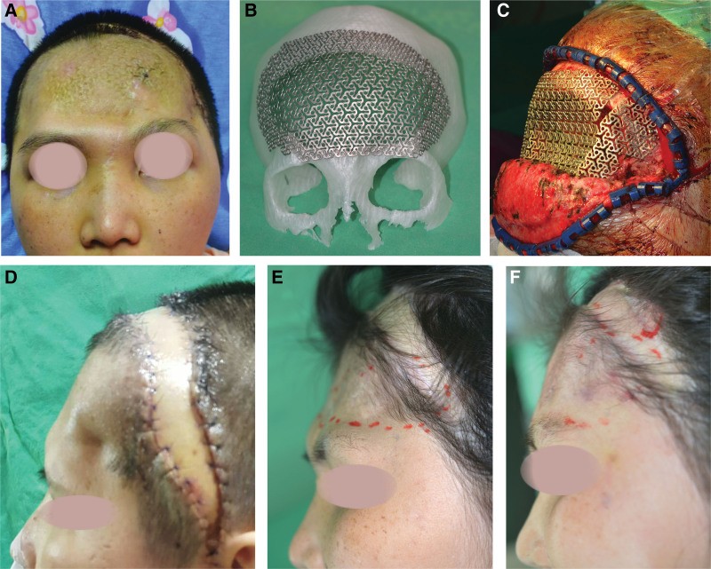
They are used mainly to replace shape in areas that do not carry much load: the skull (cranioplasty), the orbital walls, and facial contour (malar, chin, mandibular angle).
The main point of this chapter: an alloplastic implant has no blood supply and never gets one. It cannot fight infection. Its survival depends on the soft tissue covering it.
Flowchart: Alloplastic materials
- Alloplastic materials
- Metal
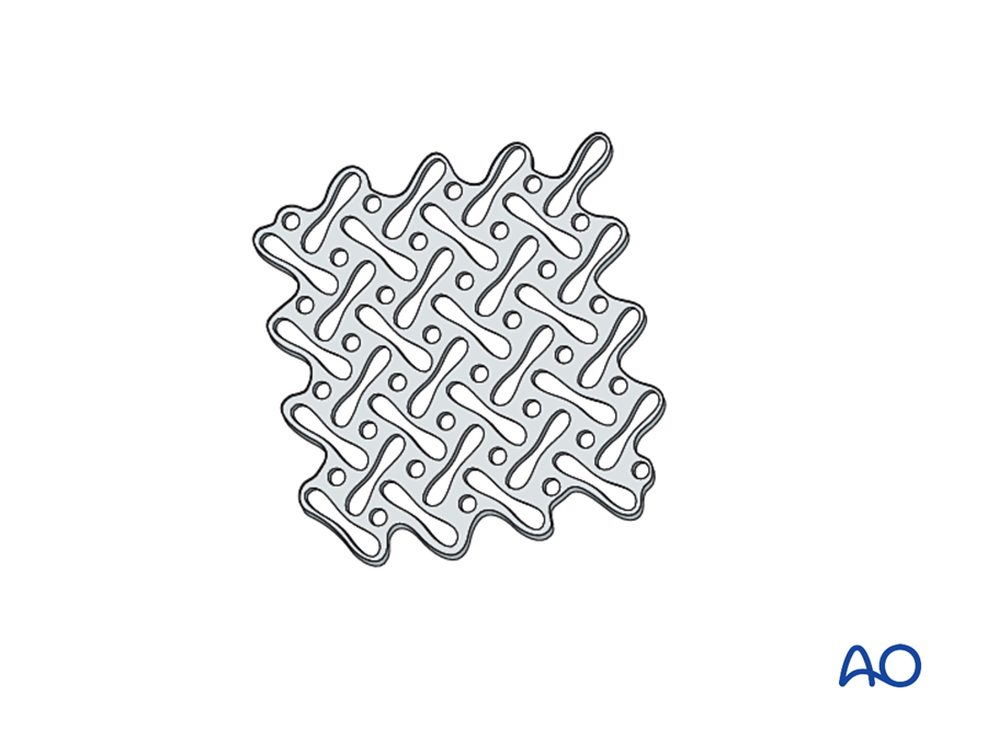
- Titaniummesh or custom plate; osseointegrates
- Polymers
- PEEKcustom implant
- PMMAbone cement
- Porous polyethylenefacial implants
- Ceramics
- Hydroxyapatite
- Calcium phosphate cement
- Bioactive glass
- Metal
12.2 The materials
| Material | Relationship with bone | How it is shaped | Advantages | Disadvantages |
|---|---|---|---|---|
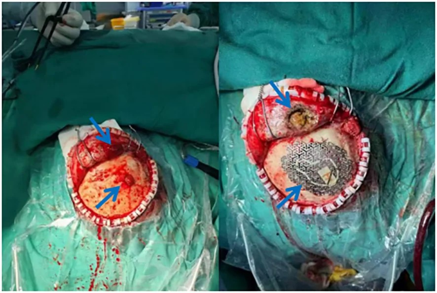
| Titanium (mesh or custom plate) | Osseointegrates | Bent in theatre, or custom-made from CT | Strong, thin, tolerates contamination better than others | Can be visible or exposed under thin scalp; artifact on CT |
| PEEK (custom implant) | No bonding; fibrous capsule | Custom-made from CT | Stiffness similar to bone; exact fit; no CT artifact | No integration; infection; cost |
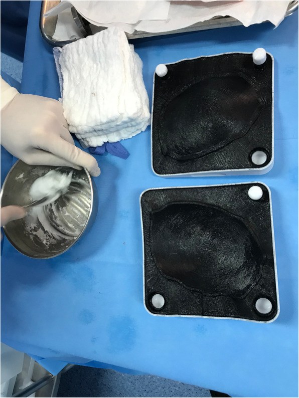
| PMMA (bone cement) | No bonding; fibrous capsule | Molded in theatre | Cheap, widely available | Heat during setting; brittle; infection |
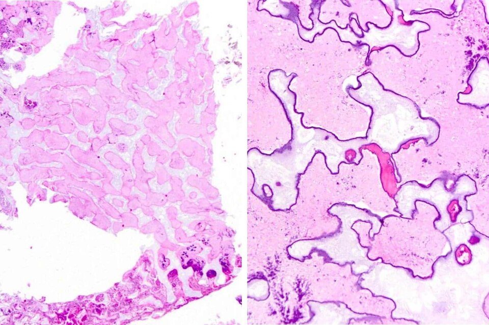
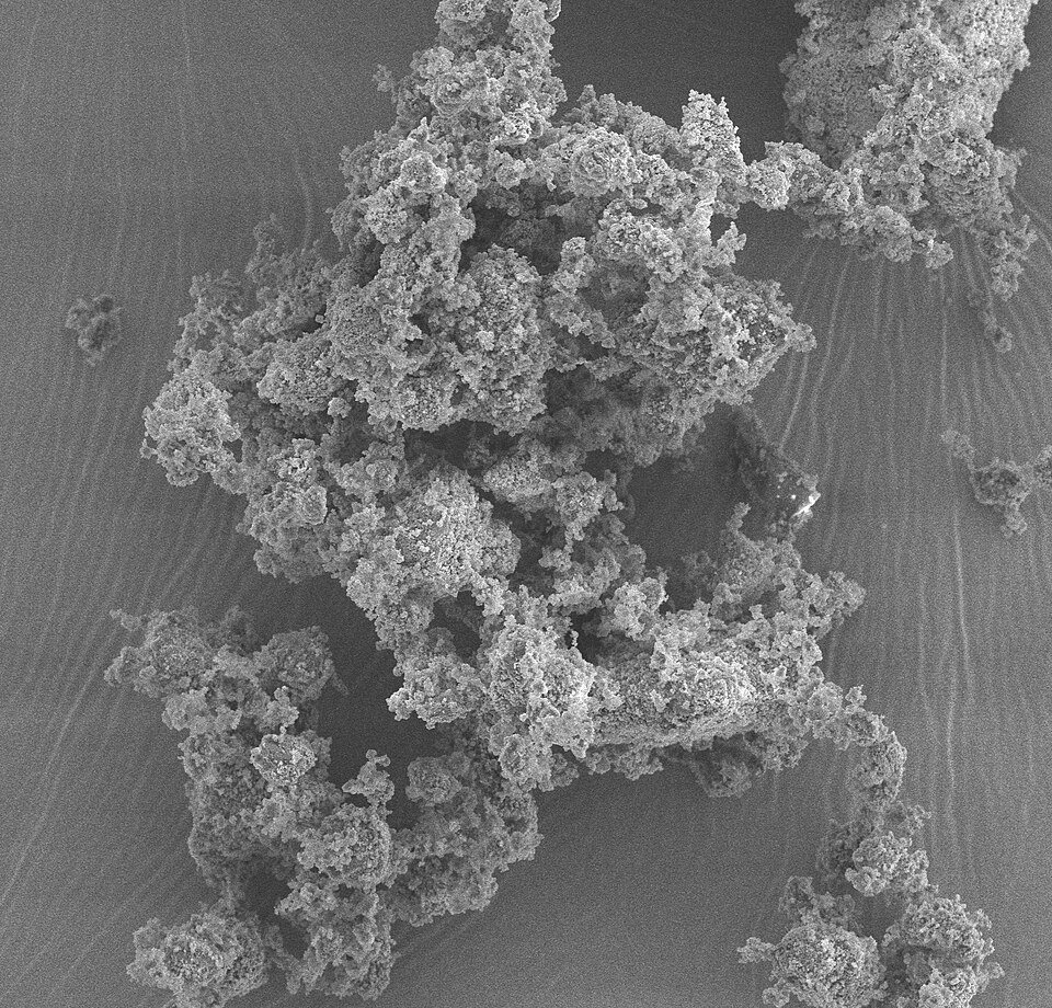
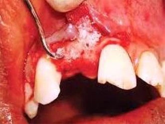
| Hydroxyapatite and calcium phosphate cement | Bonds to bone; osteoconductive for a few millimeters | Paste that sets | Good for small contour defects | Brittle; fragments in large defects; infection near sinuses |
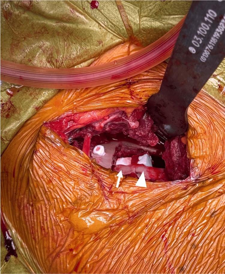
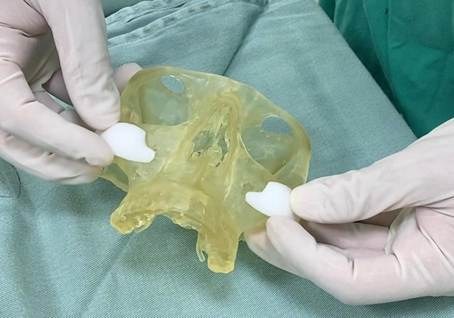
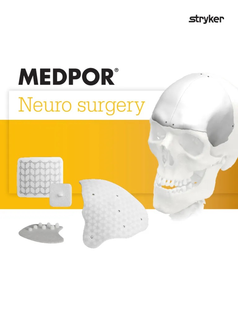
| Porous polyethylene (Medpor) | Soft tissue grows into the pores | Carved with a blade; fixed with screws | Facial contour, orbital floor | Exposure under thin cover; difficult to remove |
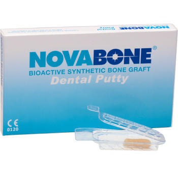
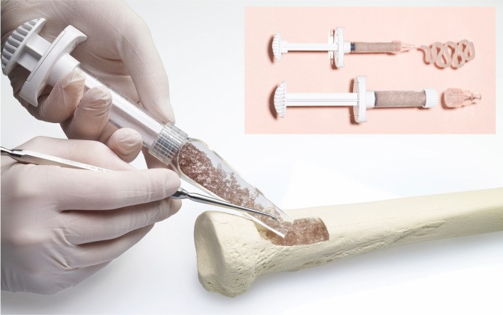
| Bioactive glass, porous ceramics | Bonds to bone | Granules, putty or blocks | Filling contained defects | Brittle; no strength |
(Gosain 2005; Rubin and Yaremchuk 1997)
12.2.1 Further details
| Property | Detail |
|---|---|
| Elastic modulus | Cortical bone 15 to 20 GPa; PEEK 3 to 4 GPa; PMMA 2 to 3 GPa; titanium about 110 GPa |
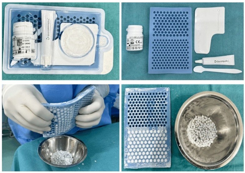
| Calcium phosphates | Hydroxyapatite (HA): very slow resorption, lasts for years. β-tricalcium phosphate (β-TCP): resorbs in months. Biphasic calcium phosphate mixes the two. Calcium sulfate: resorbs in weeks. |
| PMMA | Sets by an exothermic reaction that can exceed 70 °C; irrigate while it sets and protect the dura. Can be mixed with antibiotics. |
| Porous polyethylene | Pores of roughly 100 to 250 µm allow fibrovascular ingrowth, which fixes the implant and may reduce infection risk. |
| Bioactive glass | Silica, calcium, sodium and phosphate. In tissue fluid the surface forms a hydroxyapatite layer that bonds to bone. Has some antibacterial effect. |
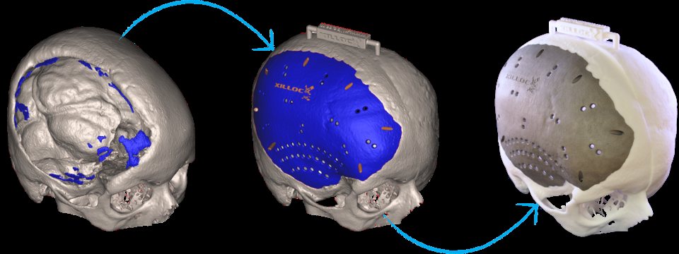
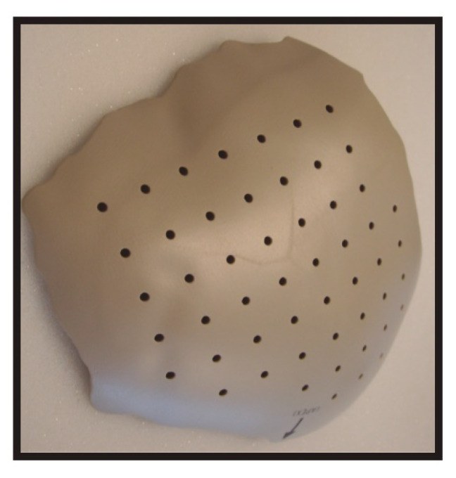
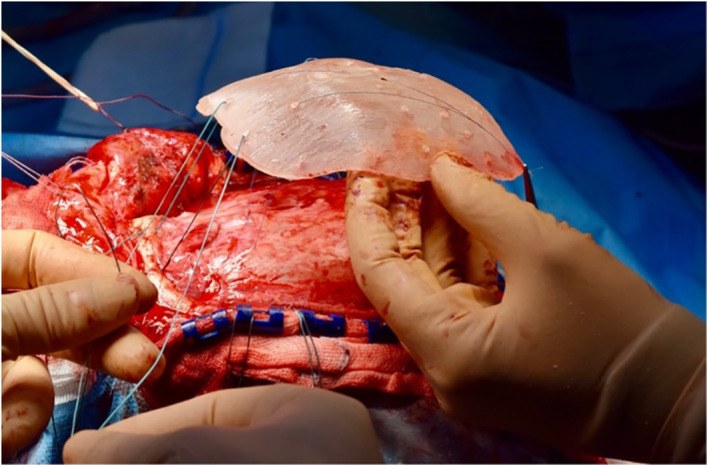
| Custom implants | CT → 3D model of the defect (mirrored from the normal side) → implant designed → milled or 3D printed in titanium or PEEK |
12.2.2 Examples of products
| Material | Examples | Company |
|---|---|---|
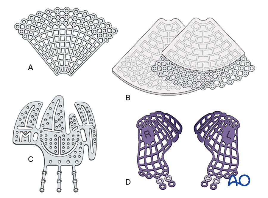
| Titanium | Mesh and plates: MatrixNEURO, MatrixORBITAL, MatrixMANDIBLE. Similar mesh, preformed orbital plates and custom implants are made by other companies. | DePuy Synthes; KLS Martin; Stryker; Medartis |
| PEEK | Custom implants milled from PEEK-Optima polymer, made to order from the CT scan | DePuy Synthes; KLS Martin; Stryker |
| PMMA | Bone cement mixed in the operating room: Palacos, Simplex P. HTR-PMI is a custom porous implant based on PMMA. | Heraeus; Stryker; Zimmer Biomet |
| Porous polyethylene | Medpor: sheets and preformed shapes for the orbit, malar area, chin, mandibular angle and ear. Medpor Titan has titanium mesh inside. Others: Su-Por, Synpor. | Stryker (first made by Porex); Poriferous; DePuy Synthes |
| Calcium phosphate cement | Norian; BoneSource; HydroSet; Mimix | DePuy Synthes; Stryker; Stryker; Zimmer Biomet |
| Hydroxyapatite | Pro Osteon (made from coral); GranuMaS (granules made in Malaysia) | Zimmer Biomet; GranuLab |
| β-tricalcium phosphate | Vitoss; chronOS; Cerasorb; Osferion | Stryker; DePuy Synthes; curasan; Olympus Terumo Biomaterials |
| Biphasic calcium phosphate | MBCP; Mastergraft | Biomatlante; Medtronic |
| Calcium sulfate | Osteoset; Stimulan (often mixed with antibiotics) | Wright Medical; Biocomposites |
| Bioactive glass | NovaBone; PerioGlas; BonAlive (S53P4) | NovaBone Products; Bonalive Biomaterials |
| Resorbable plates and mesh | LactoSorb; RapidSorb; SonicWeld Rx; Inion CPS | Zimmer Biomet; DePuy Synthes; KLS Martin; Inion |
| Polycaprolactone mesh | Osteomesh: a 3D-printed resorbable mesh for the orbital floor and burr holes | Osteopore (Singapore) |
| Silicone | Solid chin, malar and nasal implants. "Silastic" was the original trade name. | Implantech and others |
| ePTFE | Gore-Tex sheets and facial implants; no longer marketed for the face | W. L. Gore |
12.3 Cranioplasty
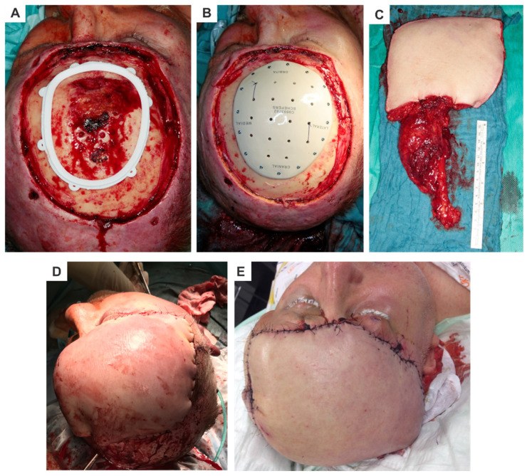
The commonest situation is a large skull defect after decompressive craniectomy. There are two choices: put back the patient's own stored bone flap, or use an alloplastic implant.
Indications for cranioplasty: protection of the brain; cosmetic contour; and treatment of the "syndrome of the trephined" (sinking skin flap syndrome): headache, dizziness, mood change and neurological deficit that improve after cranioplasty.
Timing: traditionally at least 3 months after craniectomy, and 6 to 12 months after an infection. Earlier cranioplasty is increasingly practiced in clean cases. The scalp must be healthy and relaxed enough to close without tension.
12.3.1 The stored autologous bone flap
The patient's own bone flap seems the best choice. But after storage it is a large, dead, non-vascularized cortical graft. It often resorbs.
| Study | Finding |
|---|---|
| Grant 2004: 40 children and adolescents | 50% had bone flap resorption that needed reoperation |
| Honeybul 2017: randomized trial, 64 adults | 22% of autologous bone flaps resorbed completely within 1 year. No failures with custom titanium. Total cost was not different. |
| Malcolm 2018: meta-analysis, 1,586 cranioplasties | Autologous bone had 1.9 times more reoperations than synthetic materials. Resorption in 20%. Infection rate was not different. |
So resorption is the main problem with autologous bone flaps, especially in children and young adults.
Flowchart: Cranioplasty after craniectomy
- Skull defect
- Infant under about 1 year
- Often closes by itself, or use bone graft
- Child
- Split calvarial graftstored flap resorbs in about 50%
- Adult
- Good bed
- Stored bone flapresorbs in about 20%
- Custom titanium or PEEK
- Poor bed: infected, irradiated, thin scalp, near sinus
- Improve soft tissue cover first, then autogenous bone or titanium mesh
- Good bed
- Infant under about 1 year
12.3.2 Does the choice of alloplastic material matter?
Lindner (2017) randomized 52 patients to custom hydroxyapatite or custom titanium. Infection was 7.7% versus 20.8% (not statistically significant). About 28% in each group needed reoperation.
The reoperation rate was high with both materials. In a long-term series of 312 patients, complications occurred in 23.6% (Moreira-Gonzalez 2003). This suggests that the condition of the bed is at least as important as the material.
12.3.3 Choosing the material
The choice depends on the patient's age, the condition of the bed and the size of the defect:
| Situation | Usual choice | Reason |
|---|---|---|
| Infant under about 1 year | Observe, or autogenous bone | The dura can still form bone; fixed implants restrict growth and can migrate |
| Child | Split calvarial graft; custom implant if bone is insufficient | Stored bone flaps resorb in about half of children |
| Adult, clean bed, small defect | Split calvarium, titanium mesh or cement | All work in a good bed |
| Adult, clean bed, large defect | Custom titanium or PEEK; or the stored bone flap | Stored flap resorbs completely in about 1 in 5 |
| Next to the frontal sinus | Autogenous bone; separate the sinus first (pericranial flap) | Implants and cements become infected |
| Previously infected | Delay; then autogenous bone or titanium mesh under good cover | Avoid cements and the old infected flap |
| Irradiated or thin scalp | Improve the scalp first (flap or tissue expansion), then reconstruct | An implant under poor cover will be exposed |
Based on Paige 2006; Grant 2004; Honeybul 2017; Malcolm 2018; Moreira-Gonzalez 2003; Rubin and Yaremchuk 1997.
12.4 Orbital reconstruction
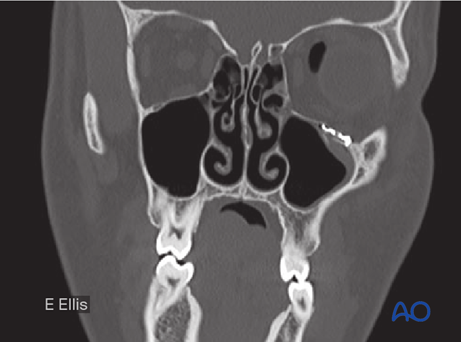
The orbital floor and medial wall do not carry load. The implant only needs to restore the shape of the orbit and hold it.
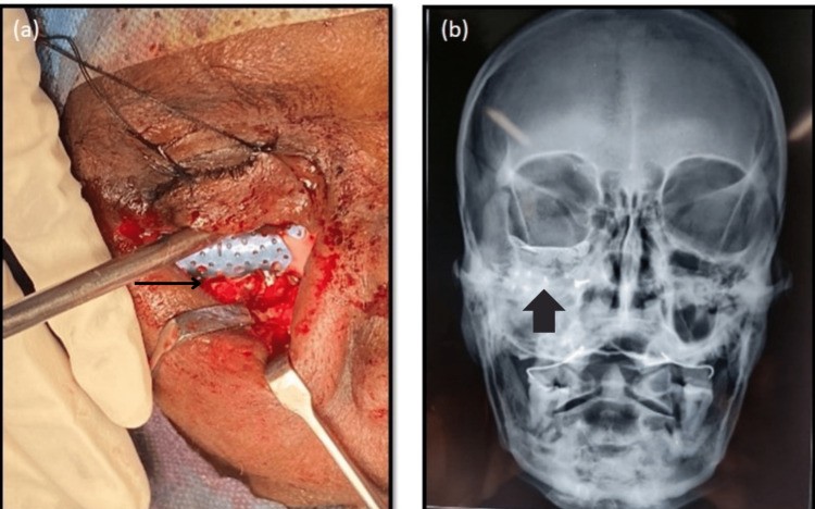
- Ellis and Tan (2003) compared titanium mesh and cranial bone graft in 58 blow-out fractures, measured on CT. Titanium mesh restored the orbital shape more accurately.
- Options: titanium mesh, porous polyethylene sheet, resorbable sheet, or bone graft.
- Bone graft is still preferred when the soft tissue cover is thin, when there is open communication with a sinus, or when the bed has been infected or irradiated.
12.5 Facial contour implants
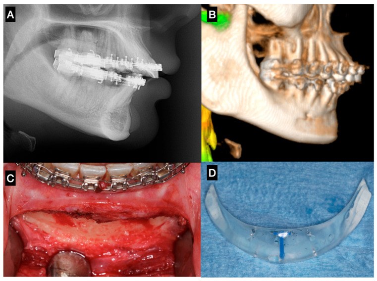
Porous polyethylene implants are widely used for the malar area, chin and mandibular angle. Yaremchuk (2003) described the principles:
- Place the implant directly on bone, under the periosteum.
- Fix it with screws, so that it does not move and there is no gap under it.
- Smooth the edges so that it cannot be felt.
- Close healthy soft tissue over it, with the incision away from the implant.
These are the same principles as for an onlay bone graft: contact, fixation and good cover.
12.6 Why implants fail
Implant failure is nearly always a problem of the bed and the soft tissue cover.
- No blood supply. Antibiotics and white cells reach bone through its vessels. They cannot reach the surface of an implant. Bacteria on the implant form a biofilm that is very hard to clear.
- Thin or tight soft tissue cover. Pressure from the implant thins the skin or mucosa until the implant is exposed.
- Communication with a contaminated space (frontal sinus, nose, mouth).
- Previous infection or radiotherapy.
An exposed bone graft can sometimes be saved, because the bed can revascularize it. An exposed implant usually has to be removed.
Implants can also fail slowly. A smooth implant that is not fixed can move on the bone. Pressure and movement cause the bone under it to resorb.
Flowchart: Order of work before placing an implant
- Control infection
- Vascularized soft tissue cover
- Tension-free closure
- Choose the material
Order of work. First control infection. Second, provide good vascularized soft tissue cover (local flap, tissue expansion or free flap). Third, close without tension. Only then choose the implant material. In a poor bed, use autogenous or vascularized tissue instead of an implant.
12.7 Summary
- Alloplastic materials replace shape, not living bone.
- Only titanium and the calcium-based materials bond to bone.
- The stored bone flap after craniectomy often resorbs; a custom implant is a valid alternative in adults.
- The soft tissue cover decides whether an implant survives.
References
- Grant GA, Jolley M, Ellenbogen RG, Roberts TS, Gruss JR, Loeser JD. Failure of autologous bone-assisted cranioplasty following decompressive craniectomy in children and adolescents. J Neurosurg 2004;100(2 Suppl Pediatrics):163–168.
- Honeybul S, Morrison DA, Ho KM, Lind CRP, Geelhoed E. A randomized controlled trial comparing autologous cranioplasty with custom-made titanium cranioplasty. J Neurosurg 2017;126:81–90.
- Malcolm JG, Mahmooth Z, Rindler RS, et al. Autologous cranioplasty is associated with increased reoperation rate: a systematic review and meta-analysis. World Neurosurg 2018;116:60–68.
- Corliss B, Gooldy T, Vaziri S, et al. Complications after in vivo and ex vivo autologous bone flap storage for cranioplasty. World Neurosurg 2016;96:510–515.
- Gosain AK; Plastic Surgery Educational Foundation DATA Committee. Biomaterials for reconstruction of the cranial vault. Plast Reconstr Surg 2005;116:663–666.
- Rubin JP, Yaremchuk MJ. Complications and toxicities of implantable biomaterials used in facial reconstructive and aesthetic surgery. Plast Reconstr Surg 1997;100:1336–1353.
- Lindner D, Schlothofer-Schumann K, Kern BC, et al. Cranioplasty using custom-made hydroxyapatite versus titanium: a randomized clinical trial. J Neurosurg 2017;126:175–183.
- Moreira-Gonzalez A, Jackson IT, Miyawaki T, Barakat K, DiNick V. Clinical outcome in cranioplasty: critical review in long-term follow-up. J Craniofac Surg 2003;14:144–153.
- Paige KT, Vega SJ, Kelly CP, et al. Age-dependent closure of bony defects after frontal orbital advancement. Plast Reconstr Surg 2006;118:977–984.
- Ellis E, Tan Y. Assessment of internal orbital reconstructions for pure blowout fractures: cranial bone grafts versus titanium mesh. J Oral Maxillofac Surg 2003;61:442–453.
- Yaremchuk MJ. Facial skeletal reconstruction using porous polyethylene implants. Plast Reconstr Surg 2003;111:1818–1827.