7.1 Introduction
This chapter describes what happens when bone is broken or cut, and how it joins again.
In plastic surgery we meet this in two ways:
- Fractures that we treat: facial fractures, hand fractures, and open tibial fractures that need flap cover.
- Osteotomies that we make ourselves: a Le Fort I osteotomy, a fibula flap cut into segments for the mandible, bone shortening in replantation. An osteotomy is a planned fracture. It heals by the same process.
Two things decide whether bone heals: stability (how much the bone ends move) and blood supply. This chapter is organized around these two.
Flowchart: Overview of bone healing
- Fracture or osteotomy
- Absolute stabilityno movement, bone ends in contact
- Primary (direct) healingno callus
- Contact healingbone ends touching
- Gap healingsmall stable gap
- Primary (direct) healingno callus
- Relative stabilitysmall controlled movement
- Secondary (indirect) healingwith callus
- 1. Hematoma → 2. Soft callus → 3. Hard callus → 4. Remodeling
- Secondary (indirect) healingwith callus
- Too much movement, or poor blood supply
- Delayed union or nonunion
- Hypertrophicneeds stability
- Atrophicneeds biology
- Delayed union or nonunion
- Absolute stabilityno movement, bone ends in contact
7.2 Two types of bone healing
Bone can heal in two ways. Which one happens depends on how much movement there is between the bone ends.
| Primary (direct) healing | Secondary (indirect) healing | |
|---|---|---|
| Condition needed | Bone ends in contact, compressed, with no movement (absolute stability) | Bone ends aligned, with a small amount of movement (relative stability) |
| What happens | Osteoclasts cut tunnels across the fracture line ("cutting cones"). Osteoblasts follow and fill the tunnels with new bone. No callus forms. | Hematoma → soft callus (cartilage) → hard callus (woven bone) → remodeling |
| On X-ray | No callus. The fracture line slowly disappears. | Callus is visible from about 3 weeks. |
| Speed | Slow (months) | Faster |
| Examples in our practice | Plated osteotomies (Le Fort I, sagittal split), fibula segments fixed with plates, lag screw fixation of a phalanx | Closed reduction of nasal and zygomatic arch fractures, K-wire fixation of fingers, condylar neck fractures treated closed, distraction osteogenesis |
7.2.1 Primary healing in detail
- Contact healing: occurs where the gap is less than about 0.01 mm and strain is under 2%. Cutting cones form at the ends of the osteons nearest the fracture. Osteoclasts at the tip of the cone cut a tunnel across the fracture line at about 50 to 100 µm per day. Osteoblasts behind them lay down lamellar bone. Union and remodeling happen at the same time.
- Gap healing: occurs where there is a stable gap of less than about 0.8 to 1 mm. The gap is first filled with lamellar bone laid across the gap, at right angles to the long axis. This bone is weak in that direction. From about 3 to 8 weeks, cutting cones cross it and replace it with osteons in the correct direction.
- Primary healing is not faster than secondary healing. Its advantage is that the fixation carries the load, so function can start early.
- Callus appearing under a compression plate means the fixation is moving ("irritation callus"). It is a warning sign.
Neither type is "better". Primary healing allows early movement because the fixation is rigid. Secondary healing is faster and stronger, but it needs the part to be protected, and it produces callus.
7.3 Stages of secondary healing
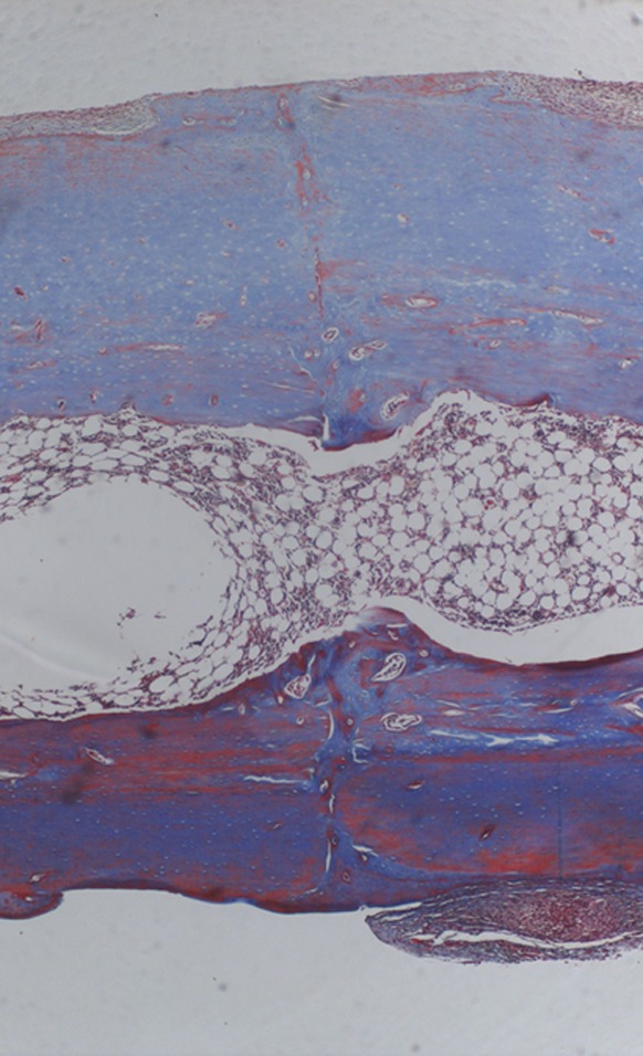
Most fractures that are not rigidly plated heal by secondary healing. It has four stages. The stages overlap.
- Hematoma and inflammation (day 0 to 5). Blood clot forms at the fracture. Platelets and inflammatory cells release growth factors. These attract stem cells from the periosteum and the marrow.
- Soft callus (day 5 to week 3). New vessels grow in. Cartilage and fibrous tissue form between the bone ends. The fracture becomes "sticky": it no longer moves easily.
- Hard callus (week 3 to 12). The cartilage is replaced by woven bone. The fracture is now clinically united: no pain and no movement.
- Remodeling (months to years). Woven bone is replaced by lamellar bone. Extra callus is removed.
Flowchart: Stages of secondary healing
- Hematoma and inflammationday 0 to 5
- Soft callusday 5 to week 3
- Hard callusweek 3 to 12
- Remodelingmonths to years
7.3.1 Cellular and molecular events
Secondary healing repeats the steps of endochondral ossification in the embryo (Gerstenfeld 2003).
| Stage | Cells | Main signals | Key points |
|---|---|---|---|
| 1. Hematoma and inflammation | Platelets, neutrophils, macrophages, lymphocytes | IL-1, IL-6, TNF-α (peak in the first 24 hours); PDGF, TGF-β from platelets; SDF-1 recruits mesenchymal stem cells (MSCs) | The hematoma is hypoxic and acidic and contains the first growth factors. Removing it delays healing (Kolar 2010). BMP-2 is required to start fracture healing (Tsuji 2006). |
| 2. Soft callus | MSCs from periosteum, endosteum and marrow; chondrocytes; fibroblasts | TGF-β2 and β3, BMP-2, 4, 7; Sox9 (cartilage); HIF-1α → VEGF | In the center (low oxygen, more movement): cartilage, collagen type II. At the periphery, under the periosteum (good oxygen, less movement): direct intramembranous bone, driven by Runx2 and Osterix. |
| 3. Hard callus | Hypertrophic chondrocytes, endothelial cells, osteoblasts, osteoclasts | Ihh and PTHrP; collagen type X; VEGF; MMP-9 and MMP-13; M-CSF and RANKL; Wnt/β-catenin | Chondrocytes hypertrophy and the matrix calcifies. Vessels invade. Cartilage is removed and replaced by woven bone. |
| 4. Remodeling | Osteoclasts and osteoblasts (coupled) | RANKL and OPG; second peak of IL-1 and TNF-α | Woven bone → lamellar bone, shaped by load (Wolff's law). |
Source of the cells. The periosteum is the most important source of progenitor cells for callus. The endosteum and marrow contribute. Surrounding muscle also supplies cells and vessels, which is one reason why muscle cover helps fracture healing.
Angiogenesis and osteogenesis are coupled. Hypoxia stabilizes HIF-1α, which increases VEGF. New vessels bring oxygen, nutrients and osteoprogenitor cells. A specific type of bone capillary (type H) is linked to osteoprogenitors (Kusumbe 2014).
Times are for an adult long bone. Facial bones and children heal faster. (Einhorn and Gerstenfeld 2015)
What this means in practice. A displaced nasal or zygomatic fracture can be reduced by simple elevation only in the first 1 to 2 weeks, before soft callus fixes it in the wrong position. After that, an osteotomy is needed. In the hand, K-wires are removed at 3 to 4 weeks, when there is clinical union, even though the fracture line is still visible on X-ray.
7.4 Stability and strain
The amount of movement at the fracture decides the type of healing. The reason is strain.
Strain is the amount of movement at the fracture divided by the width of the gap:
strain = movement ÷ gap width
Each tissue tolerates a different amount of strain (Perren 2002):
- Bone: up to about 2%
- Cartilage: up to about 10%
- Granulation tissue: up to 100%
So if strain is under 2%, bone can form directly (primary healing). If strain is between 2% and 10%, cartilage forms first, makes the fracture stiffer, and then bone can form (secondary healing). If strain is too high, no tissue can bridge the gap and the result is nonunion. These numbers are a guide, not exact limits.
Flowchart: Strain decides the tissue that forms
- Strain = movement ÷ gap width
- Under 2%
- Bone forms directlyprimary healing
- 2% to 10%
- Cartilage, then bonesecondary healing
- 10% to 100%
- Granulation tissue onlydelayed healing
- Over 100%
- No tissue bridgesnonunion
- Under 2%
7.4.1 Other mechanobiology theories
| Author | Main idea |
|---|---|
| Pauwels (1960) | Type of mechanical stimulus decides the tissue: hydrostatic pressure → cartilage; shear and stretch → fibrous tissue; bone forms only when these soft tissues have made the site stable. |
| Perren (1979) | Interfragmentary strain theory: a tissue can only form if it tolerates the strain present (bone 2%, cartilage 10%, granulation tissue 100%). |
| Carter (1988) | Combines hydrostatic stress, strain and blood supply: low stress and good vascularity → bone; compression with poor vascularity → cartilage; high tensile strain → fibrous tissue. |
| Claes and Heigele (1999) | Quantified the limits: strain under about 5% with low pressure → intramembranous bone; strain under about 15% with compressive pressure → endochondral bone; larger values → fibrous tissue. |
- Some movement stimulates callus. In experimental osteotomies, axial movement of about 0.2 to 1 mm in a gap of 1 to 2 mm gave the best callus.
- Axial (compressive) movement is tolerated better than shear.
- Large gaps heal poorly even when they are stable. A gap over about 2 mm delays healing.
- Callus increases the diameter of the bone at the fracture. Bending stiffness rises with the fourth power of the radius, so a small amount of peripheral callus stabilizes the fracture efficiently.
Worked examples:
| Situation | Gap | Movement | Strain | Result |
|---|---|---|---|---|
| 1. Osteotomy compressed under a plate (fibula segment, Le Fort I, lag-screwed phalanx) | 0.2 mm | 0 | 0% | Primary healing |
| 2. Fracture held by K-wires or a splint | 1.5 mm | 0.12 mm | 8% | Secondary healing with callus |
| 3. Tightly reduced osteotomy with slightly loose fixation | 0.2 mm | 0.12 mm | 60% | Delayed union or nonunion |
| 4. Comminuted zone bridged by a long plate | 8 mm (shared by many small gaps) | 0.5 mm | 6% | Secondary healing with callus |
| 5. Failed fixation | 1 mm | 1.6 mm | 160% | Nonunion |
Important point. Compare examples 2 and 3. The movement is the same (0.12 mm). In a 1.5 mm gap this gives 8% strain, and the fracture heals with callus. In a 0.2 mm gap it gives 60% strain, and it does not heal. This means a tightly reduced osteotomy with slightly loose fixation is the worst situation. A tight osteotomy must be fixed rigidly.
Evidence from fibula flap reconstruction
This rule has been measured in free fibula reconstruction of the jaw. On follow-up CT scans (Swendseid 2020):
- When there was a gap of 1 mm or more between bone ends, 79% did not unite completely.
- When the bone ends were in contact, only 8% did not unite completely.
- Nonunion on CT was followed by more wound complications (40% versus 19%).
The type of plate was less important. In 117 fibula flaps, miniplates and reconstruction plates had the same nonunion rate (14% and 13%) (Robey 2008). Good bone contact matters more than the plate.
7.5 Blood supply of bone
Stability is the first requirement. The second is blood supply. The cortex of a long bone has two blood supplies:
- The endosteal (medullary) supply from the nutrient artery. It supplies the marrow and the inner two-thirds of the cortex.
- The periosteal supply from the periosteum and attached muscle. It supplies the outer one-third of the cortex.
Flowchart: Blood supply of the cortex
- Blood supply of long bone cortex
- Endosteal (medullary)nutrient artery
- Inner 2/3 of cortex
- Lost in: segmental osteotomy, reaming
- Periostealperiosteum and muscle
- Outer 1/3 of cortex
- Lost in: stripping, open fracture
- Endosteal (medullary)nutrient artery
Injury and surgery can remove one or both. There are four possible situations:
| Situation | Endosteal supply | Periosteal supply | Result |
|---|---|---|---|
| Intact bone | Present | Present | Whole cortex alive |
| Periosteum stripped (wide exposure, plate pressed on bone, open fracture) | Present | Lost | Outer cortex at risk; the cells that form external callus are lost |
| Medullary supply lost (segment of a fibula flap after osteotomy; reamed canal; middle fragment of a segmental fracture) | Lost | Present | Bone survives on the periosteum and its muscle cuff |
| Both lost (free fragment; non-vascularized bone graft) | Lost | Lost | Bone cells die; the bone acts as a scaffold only |
(Rhinelander 1974)
- Periosteum stripped: happens with wide exposure for plating. Keep periosteal stripping to the minimum needed.
- Medullary supply lost: this is the situation of a fibula flap segment after osteotomy. The segment survives only on its periosteum. This is why we never strip the periosteum from a fibula flap.
- Both lost: this is a non-vascularized bone graft.
7.6 Soft tissue cover
The periosteum and surrounding muscle supply blood to bone. In an open fracture this soft tissue is lost. Exposed bone without soft tissue cover does not heal, and it becomes infected. Flap cover brings back a blood supply.
7.6.1 Classification of open fractures (Gustilo and Anderson)
| Type | Description | Infection rate reported by Gustilo |
|---|---|---|
| I | Wound under 1 cm, clean, simple fracture | 0 to 2% |
| II | Wound 1 to 10 cm, moderate soft tissue damage, no flaps or avulsion | 2 to 7% |
| IIIA | High-energy injury or extensive laceration, but bone can still be covered by local soft tissue | 4% |
| IIIB | Extensive soft tissue loss with periosteal stripping and exposed bone. Needs flap cover. | 52% |
| IIIC | Any open fracture with an arterial injury that needs repair | 42% |
Gustilo and Anderson 1976; Gustilo, Mendoza and Williams 1984. The type is decided after debridement, not at first inspection.
Principles of management of a Gustilo IIIB fracture (orthoplastic approach): early antibiotics; debridement by senior orthopedic and plastic surgeons together; skeletal stabilization; temporary dressing (negative pressure); definitive soft tissue cover as early as possible, within 72 hours where the patient and wound allow (British BOAST standard); bone reconstruction later if there is a defect.
Choice of flap by level in the leg: proximal third: gastrocnemius muscle flap; middle third: soleus flap or local fasciocutaneous flap; distal third: free flap (or propeller or reverse sural flap in selected cases).
Godina studied 532 patients with limb trauma treated by free flap. He compared early and late flap cover:
| Flap within 72 hours | 72 hours to 3 months | After 3 months | |
|---|---|---|---|
| Flap failure | 0.75% | 12% | 9.5% |
| Infection | 1.5% | 17.5% | 6% |
| Time to bone union | 6.8 months | 12.3 months | 29 months |
Early cover gave less infection and faster bone union. Later studies have questioned the exact 72-hour limit, but the principle of early cover remains. In the "fix and flap" series from Leeds (84 severe open tibial fractures treated by orthopedic and plastic surgeons together), limb salvage was 95% (Gopal 2000).
Local data (USM). Yusof and Halim (2012) reported 11 infected Gustilo IIIB open tibial fractures treated in stages: repeated debridement, flap cover, then bone reconstruction. Infection resolved in all 11. Nine united, at a mean of 15 months. Compare this with 6.8 months in Godina's early group: delay and infection make union much slower.
7.7 Fixation of the facial skeleton
Plastic surgeons fix bone most often in the face and the hand.
7.7.1 Mandible
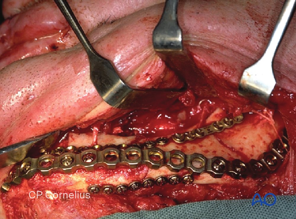
When the mandible is loaded during biting, the upper border (near the teeth) is pulled apart. This is the tension zone. The lower border is pushed together. This is the compression zone.
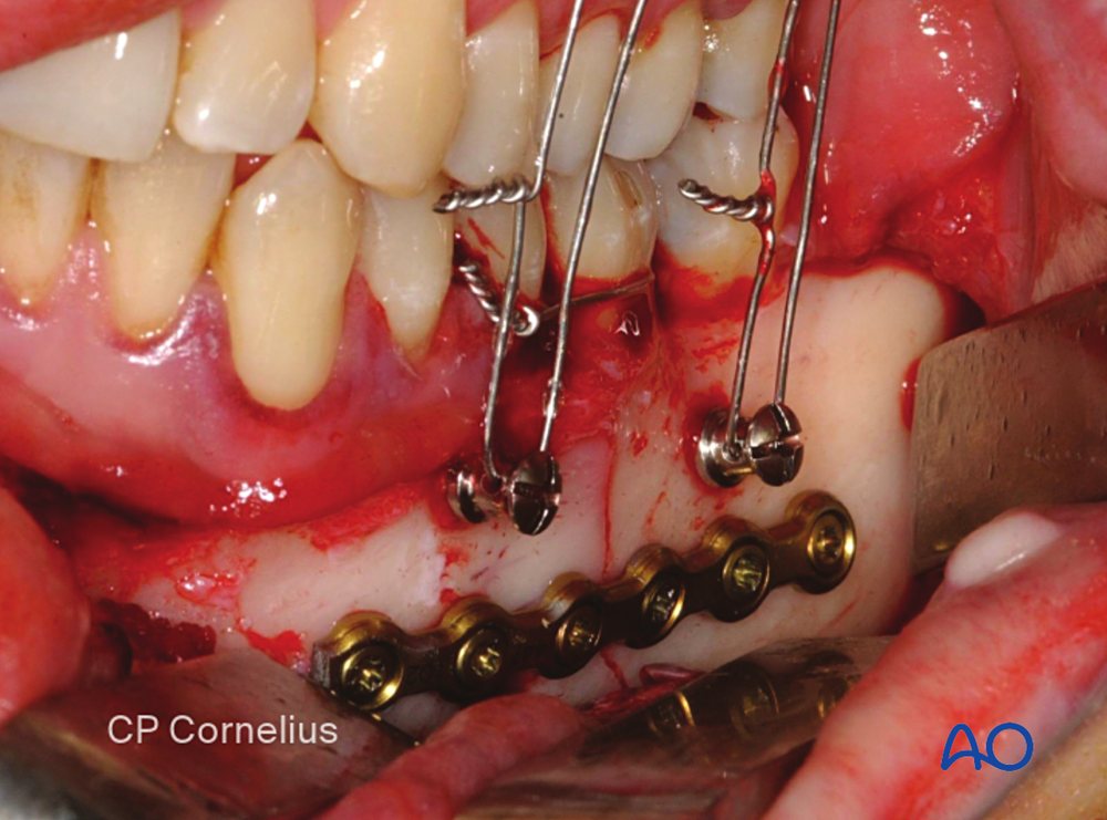
Champy showed that a small plate placed along the tension zone is enough to hold a simple fracture, because the bone itself carries the compression at the lower border. These positions are called Champy's lines of osteosynthesis: one plate at the angle (on the external oblique ridge), one plate in the body (below the roots), and two plates at the symphysis (because of torsion).
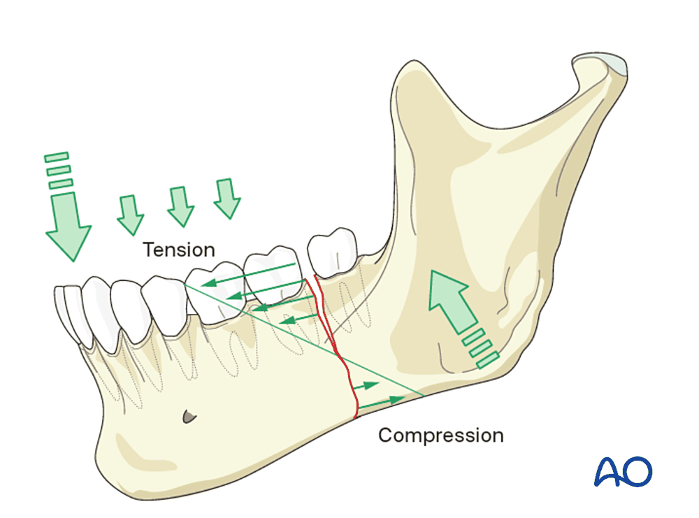
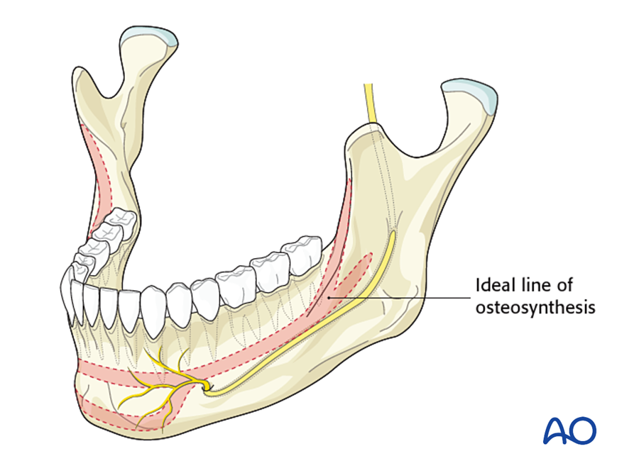
(Michelet 1973; Champy 1978; Ellis 1999)
This gives two types of fixation:
Flowchart: Choosing fixation in the mandible
- Mandibular fracture or defect
- Bone can share the loadsimple fracture, good bone
- Load-sharing fixationminiplate on the tension zone (Champy)
- Bone cannot share the loadcomminuted, atrophic, infected, defect
- Load-bearing fixationreconstruction plate
- Bone can share the loadsimple fracture, good bone
7.7.2 Midface
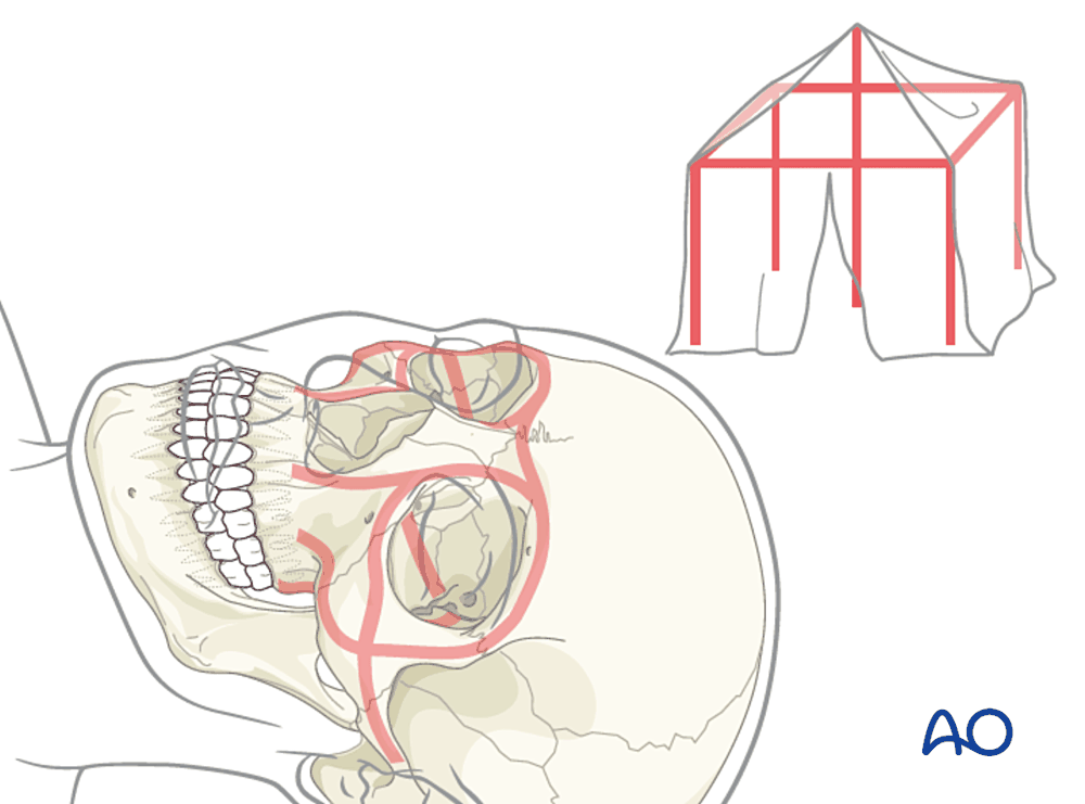
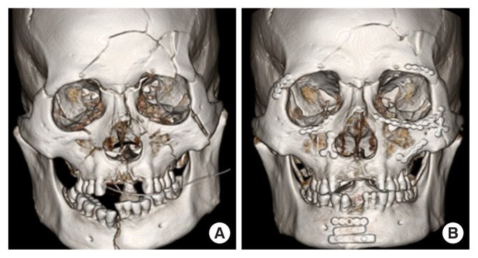
The midface is made of thick vertical pillars of bone called buttresses, with thin bone between them. The buttresses carry the chewing force up to the skull base. There are three on each side: nasomaxillary, zygomaticomaxillary and pterygomaxillary (Manson 1980).
- In midface fractures, we plate the buttresses. This restores the height and projection of the face.
- If part of a buttress is missing, it is replaced with a bone graft at the same operation (Gruss and Mackinnon 1986).
- The thin walls (orbital floor, sinus wall) do not carry load. They only need a thin sheet to restore their shape: bone graft, titanium mesh or a polymer sheet.
7.7.3 The skull in young children
The dura of an infant can form new bone. Small defects of the skull in infants can close by themselves. This ability is lost early. In 81 children after fronto-orbital advancement, bone gaps closed if surgery was before about 9 to 11 months of age, and stayed open if surgery was later (Paige 2006). After this age, skull defects need a bone graft.
7.8 Fixation in the hand
In the hand the main problem after a fracture is usually stiffness, not nonunion. The flexor and extensor tendons lie directly on the periosteum of the phalanges. Callus, plates and surgical dissection all cause the tendons to stick to the bone.
Page and Stern (1998) reviewed 105 metacarpal and phalangeal fractures fixed with plates:
- 36% had a major complication (stiffness, nonunion, plate prominence, infection, tendon rupture).
- Only 11% of plated phalangeal fractures regained good movement (total active motion over 220°).
So in the hand we choose the smallest fixation that still allows early movement: K-wires or lag screws where possible, plates only when needed.
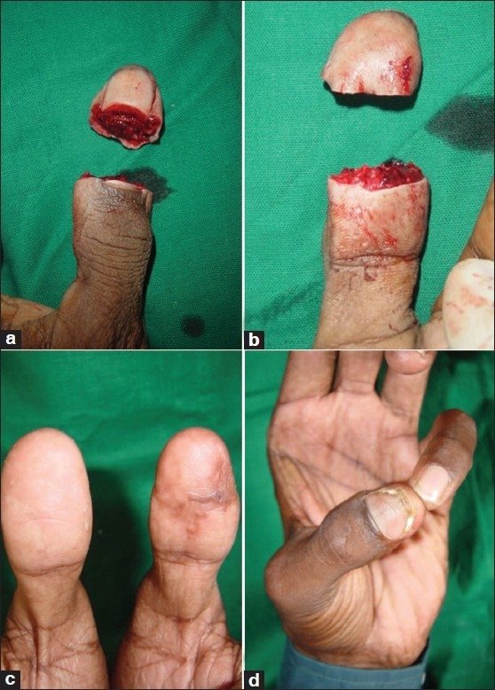
Replantation. In replantation the bone is shortened by a few millimeters before fixation. This is done on purpose. It allows the vessels, nerves and skin to be repaired without tension and often without vein grafts.
7.9 Factors that delay bone healing
So far we have covered what bone needs to heal. This section lists what interferes with it. It is useful to separate patient factors from surgical factors, because the surgical factors are under our control.
Patient factors
- Smoking: 2.2 times the risk of delayed union or nonunion (meta-analysis of 7,516 procedures; Pearson 2016)
- Diabetes, malnutrition, vitamin D deficiency
- Steroids, chemotherapy
- Old age, vascular disease
- Previous radiotherapy (see below)
Surgical factors
- Heat: bone cells die at 47 °C for one minute (Eriksson and Albrektsson 1983). Always irrigate when sawing or drilling.
- Too much periosteal stripping
- A gap between bone ends, or loose fixation
- Wound closed under tension, or poor soft tissue cover
- Infection: in the mouth, the mucosa over a plate must be closed watertight
| Factor | Mechanism | Practical point |
|---|---|---|
| Smoking | Nicotine: vasoconstriction, inhibits osteoblasts and angiogenesis. Carbon monoxide: reduces oxygen delivery. | Stop at least 4 weeks before elective bone surgery |
| NSAIDs | Inhibit COX-2 and prostaglandin E2, which are needed in the inflammatory and early callus stages | Animal evidence is clear; human evidence is mixed. Avoid prolonged high doses after bone surgery. |
| Corticosteroids | Reduce osteoblast differentiation; increase osteocyte apoptosis | |
| Diabetes | Fewer and less active MSCs; microangiopathy; advanced glycation end-products in collagen; higher infection rate | Control glucose perioperatively |
| Malnutrition | Low protein, vitamin C (collagen), vitamin D and calcium | Check albumin and vitamin D in nonunion |
| Bisphosphonates, denosumab | Suppress osteoclasts and remodeling | Risk of medication-related osteonecrosis of the jaw (MRONJ) after dental extraction or jaw surgery |
| Infection | Bacterial biofilm on dead bone and implants; inflammation causes bone resorption | Debride dead bone; stable fixation; soft tissue cover |
Irradiated bone
Radiotherapy damages bone permanently. Marx (1983) described irradiated tissue with "three H's":
- Hypovascular: small vessels are narrowed and reduced.
- Hypocellular: osteoblasts and their stem cells are reduced.
- Hypoxic: oxygen level in the tissue is low.
Irradiated bone cannot repair itself after injury, such as a tooth extraction or an osteotomy. This leads to osteoradionecrosis. A non-vascularized bone graft will not survive in an irradiated bed, because there are no vessels to grow into it. Bone placed here must bring its own blood supply: a vascularized bone flap.
Two theories of osteoradionecrosis (ORN):
| Theory | Mechanism | Treatment that follows from it |
|---|---|---|
| Marx (1983): hypoxic, hypovascular, hypocellular tissue | Radiation → endarteritis → tissue hypoxia → tissue breakdown → chronic non-healing wound. Infection is secondary. | Hyperbaric oxygen with surgery (Marx protocol). Later trials have not confirmed a clear benefit of hyperbaric oxygen alone. |
| Delanian (2004): radiation-induced fibroatrophic process | Radiation → free radicals and endothelial injury → abnormal fibroblast activation → fibrosis → fragile, poorly vascularized tissue | Pentoxifylline, tocopherol (vitamin E) and clodronate (PENTOCLO) |
| Notani class | Extent of ORN in the mandible | Usual treatment |
|---|---|---|
| I | Alveolar bone only | Conservative: oral hygiene, antibiotics, pentoxifylline and tocopherol, limited sequestrectomy |
| II | Alveolar bone and mandible above the inferior alveolar canal | Sequestrectomy or marginal resection, often with vascularized soft tissue |
| III | Below the canal, or with pathological fracture or skin fistula | Segmental resection and free vascularized bone flap |
Risk rises with dose (especially above 60 Gy), mandible more than maxilla, and dental extraction after radiotherapy.
7.10 Delayed union and nonunion
Healing may be slow, or it may fail.
- Delayed union: the fracture is healing, but more slowly than expected.
- Nonunion: healing has stopped, and the fracture will not unite without further treatment.
Working definition (FDA): nonunion is a fracture at least 9 months old that has shown no progression of healing for 3 consecutive months. The time varies with the bone: a mandible or a metacarpal is expected to unite much sooner than a tibia.
7.10.1 Classification (Weber and Cech)
| Group | Type | Appearance | Problem |
|---|---|---|---|
| Viable (hypervascular) | Hypertrophic, "elephant foot" | Abundant callus | Instability |
| Slightly hypertrophic, "horse hoof" | Some callus | Instability | |
| Oligotrophic | No callus, but bone ends are alive | Displacement or poor contact | |
| Non-viable (avascular) | Torsion wedge | A dead intermediate fragment, united on one side only | Poor blood supply |
| Comminuted | Several dead fragments | Poor blood supply | |
| Defect | Bone loss | Gap | |
| Atrophic | Thin, tapered, sclerotic ends | Poor blood supply; end result of the above |
Infected nonunion is considered separately, because infection must be treated first.
7.10.2 Assessment
- History: injury, previous surgery, infection, smoking, diabetes, drugs, radiotherapy.
- Examination: pain, abnormal movement, soft tissue, sinus, neurovascular status.
- Blood tests: white cell count, ESR, CRP; vitamin D, calcium, glucose, albumin.
- Imaging: X-rays; CT to confirm bridging. The RUST score (Radiographic Union Score for Tibial fractures) grades each of 4 cortices from 1 to 3; total 4 (not healed) to 12 (healed) (Whelan 2010).
- At surgery: multiple deep tissue samples for culture.
The X-ray appearance shows what was missing, and so what treatment is needed:
Hypertrophic nonunion. A lot of callus, wide bone ends, but a line remains. The biology is good. The problem is too much movement.
Treatment: more stable fixation. Bone graft is usually not needed.
Atrophic nonunion. No callus, thin tapered bone ends. The problem is poor biology: poor blood supply and no bone-forming activity.
Treatment: remove dead bone until the ends bleed, add bone graft, give stable fixation, and provide good soft tissue cover (often a flap).
Figure 7.11. Hypertrophic and atrophic nonunion.
Flowchart: Approach to nonunion
- Fracture not uniting
- Check: infection, fixation, blood supply, patient factors
- Hypertrophiccallus present
- Improve stabilityrevise fixation
- Atrophicno callus
- Improve biologydebride, bone graft, flap cover, stable fixation
- Infected
- Debride, treat infection, then reconstruct in stages
- Hypertrophiccallus present
- Check: infection, fixation, blood supply, patient factors
The diamond concept
Giannoudis (2007) summarized the requirements for bone healing as four items, called the diamond concept:
- Mechanical stability (this chapter)
- Osteogenic cells
- Growth factors (osteoinductive signals)
- Scaffold (osteoconductive matrix)
All of these must be in a well-vascularized bed. Items 2 to 4 are what a bone graft provides.
References
- Perren SM. Evolution of the internal fixation of long bone fractures. J Bone Joint Surg Br 2002;84:1093–1110.
- Einhorn TA, Gerstenfeld LC. Fracture healing: mechanisms and interventions. Nat Rev Rheumatol 2015;11:45–54.
- Savaridas T, Wallace RJ, et al. The development of a novel model of direct fracture healing in the rat. Bone Joint Res 2012;1:289–296.
- Swendseid B, Kumar A, Sweeny L, et al. Natural history and consequences of nonunion in mandibular and maxillary free flaps. Otolaryngol Head Neck Surg 2020;163:956–962.
- Robey AB, Spann ML, McAuliff TM, et al. Comparison of miniplates and reconstruction plates in fibular flap reconstruction of the mandible. Plast Reconstr Surg 2008;122:1733–1738.
- Paige KT, Vega SJ, Kelly CP, et al. Age-dependent closure of bony defects after frontal orbital advancement. Plast Reconstr Surg 2006;118:977–984.
- Greenwald JA, Mehrara BJ, Spector JA, et al. Biomolecular mechanisms of calvarial bone induction: immature versus mature dura mater. Plast Reconstr Surg 2000;105:1382–1392.
- Michelet FX, Deymes J, Dessus B. Osteosynthesis with miniaturized screwed plates in maxillo-facial surgery. J Maxillofac Surg 1973;1:79–84.
- Champy M, Loddé JP, Schmitt R, Jaeger JH, Muster D. Mandibular osteosynthesis by miniature screwed plates via a buccal approach. J Maxillofac Surg 1978;6:14–21.
- Ellis E. Treatment methods for fractures of the mandibular angle. Int J Oral Maxillofac Surg 1999;28:243–252.
- Manson PN, Hoopes JE, Su CT. Structural pillars of the facial skeleton. Plast Reconstr Surg 1980;66:54–62.
- Gruss JS, Mackinnon SE. Complex maxillary fractures: role of buttress reconstruction and immediate bone grafts. Plast Reconstr Surg 1986;78:9–22.
- Page SM, Stern PJ. Complications and range of motion following plate fixation of metacarpal and phalangeal fractures. J Hand Surg Am 1998;23:827–832.
- Rhinelander FW. Tibial blood supply in relation to fracture healing. Clin Orthop Relat Res 1974;105:34–81.
- Godina M. Early microsurgical reconstruction of complex trauma of the extremities. Plast Reconstr Surg 1986;78:285–292.
- Gopal S, Majumder S, Batchelor AG, et al. Fix and flap: the radical orthopaedic and plastic treatment of severe open fractures of the tibia. J Bone Joint Surg Br 2000;82:959–966.
- Yusof NM, Halim AS. Outcomes of infected grade IIIB open tibial fractures. Singapore Med J 2012;53:591–594.
- Marx RE. Osteoradionecrosis: a new concept of its pathophysiology. J Oral Maxillofac Surg 1983;41:283–288.
- Pearson RG, Clement RGE, Edwards KL, Scammell BE. Do smokers have greater risk of delayed and non-union after fracture, osteotomy and arthrodesis? BMJ Open 2016;6:e010303.
- Eriksson AR, Albrektsson T. Temperature threshold levels for heat-induced bone tissue injury. J Prosthet Dent 1983;50:101–107.
- Giannoudis PV, Einhorn TA, Marsh D. Fracture healing: the diamond concept. Injury 2007;38 Suppl 4:S3–S6.
- Adani R, Marcoccio I, et al. Indian J Plast Surg 2011 (source of the replantation photographs; CC BY-NC-SA 3.0).
- Gerstenfeld LC, Cullinane DM, Barnes GL, Graves DT, Einhorn TA. Fracture healing as a post-natal developmental process. J Cell Biochem 2003;88:873–884.
- Marsell R, Einhorn TA. The biology of fracture healing. Injury 2011;42:551–555.
- Kolar P, Schmidt-Bleek K, Schell H, et al. The early fracture hematoma and its potential role in fracture healing. Tissue Eng Part B Rev 2010;16:427–434.
- Tsuji K, Bandyopadhyay A, Harfe BD, et al. BMP2 activity, although dispensable for bone formation, is required for the initiation of fracture healing. Nat Genet 2006;38:1424–1429.
- Kusumbe AP, Ramasamy SK, Adams RH. Coupling of angiogenesis and osteogenesis by a specific vessel subtype in bone. Nature 2014;507:323–328.
- Claes LE, Heigele CA. Magnitudes of local stress and strain along bony surfaces predict the course and type of fracture healing. J Biomech 1999;32:255–266.
- Gustilo RB, Anderson JT. Prevention of infection in the treatment of one thousand and twenty-five open fractures of long bones. J Bone Joint Surg Am 1976;58:453–458.
- Gustilo RB, Mendoza RM, Williams DN. Problems in the management of type III (severe) open fractures. J Trauma 1984;24:742–746.
- Delanian S, Lefaix JL. The radiation-induced fibroatrophic process: therapeutic perspective via the antioxidant pathway. Radiother Oncol 2004;73:119–131.
- Notani K, Yamazaki Y, Kitada H, et al. Management of mandibular osteoradionecrosis corresponding to the severity of osteoradionecrosis and the method of radiotherapy. Head Neck 2003;25:181–186.
- Ruggiero SL, Dodson TB, Fantasia J, et al. AAOMS position paper on medication-related osteonecrosis of the jaw: 2014 update. J Oral Maxillofac Surg 2014;72:1938–1956.
- Weber BG, Cech O. Pseudarthrosis. Bern: Hans Huber; 1976.
- Whelan DB, Bhandari M, Stephen D, et al. Development of the radiographic union score for tibial fractures. J Trauma 2010;68:629–632.
- Hak DJ, Fitzpatrick D, Bishop JA, et al. Delayed union and nonunions: epidemiology, clinical issues, and financial aspects. Injury 2014;45 Suppl 2:S3–S7.