6.1 Introduction
Bone is renewed throughout life. Old bone is removed and new bone is laid down in its place. In an adult, most of the skeleton is replaced over about ten years.
This renewal does three jobs: it repairs small cracks before they join into a fracture, it adapts the bone to the loads placed on it, and it releases calcium when the blood needs it.
The same processes decide what happens after a reconstruction. A fibula flap thickens when it is loaded. Bone under a rigid plate becomes thinner. An onlay graft that carries no load is slowly resorbed. And drugs that suppress remodeling, which many of our older patients take, change how the jaw heals.
Flowchart: Three ways bone changes after it has formed
- Changes in formed bone
- Modelingchanges size and shape
- Formation and removal on different surfacesgrowth in width; adaptation to load
- Remodelingrenews bone in the same place
- Removal followed by formation at the same siterepair of microdamage; calcium release
- Repairrestores continuity after injury
- Fracture, osteotomy, graft junctionends with remodeling
- Modelingchanges size and shape
6.2 Modeling, remodeling and repair
| Process | What happens | Example |
|---|---|---|
| Modeling | Bone is added on one surface and removed from another. The bone changes size or shape. Formation and removal are not linked at one site. | Growth of a bone in width; a loaded fibula flap getting thicker |
| Remodeling | A small volume of bone is removed and then replaced at the same site. Size and shape stay the same. | Renewal of cortex by new osteons; removal of microdamage |
| Repair | New tissue forms to restore continuity after injury, and is then remodeled | Fracture callus; healing at the end of a graft |
In the adult skeleton, remodeling is the main process. Modeling is the main process during growth, but it also continues in adults when the load on a bone changes.
6.3 The remodeling cycle
Remodeling is carried out by a team of cells called the basic multicellular unit (BMU): osteoclasts in front, osteoblasts behind, with a capillary and supporting cells between them. In cancellous bone, the team works under a canopy of flat lining cells that seals off a small space from the marrow, the bone remodeling compartment (Hauge 2001).
Flowchart: The remodeling cycle at one site
- Activationa signal recruits osteoclast precursors
- Resorptionosteoclasts remove bone; about 2 to 4 weeks
- Reversalthe surface is cleaned and prepared; osteoblasts arrive
- Formationosteoblasts fill the cavity with osteoid; about 3 to 4 months
- Mineralization and restlining cells cover the new surface
Two features of this cycle matter clinically:
- Removal is fast and formation is slow. Osteoclasts remove a volume of bone in weeks; osteoblasts take months to refill it. At any moment, part of the skeleton consists of cavities that have been dug but not yet filled. This is called the remodeling space. When remodeling speeds up, for example after a fracture, the remodeling space grows and the bone becomes weaker for a time.
- The result depends on balance. If each cavity is refilled completely, bone mass stays the same. If osteoblasts refill a little less than osteoclasts removed, bone is lost with every cycle. In aging and after the menopause, this small deficit is repeated across millions of sites.
6.3.1 Cortical and cancellous remodeling
In cortical bone, the BMU moves through the bone as a tunnel: a cutting cone of osteoclasts followed by a closing cone of osteoblasts, which leaves behind a new osteon. In cancellous bone, the BMU works on the surface of a trabecula and digs a shallow trench that is then refilled, forming a "hemi-osteon" (Parfitt 1994).
When cancellous remodeling is too active, osteoclasts can cut right through a thin trabecula. That connection is lost for good; osteoblasts need a surface to build on and cannot bridge the gap. When cortical remodeling is too active, the cortex becomes porous.
6.3.2 Turnover and balance are different
High turnover means that remodeling is happening at many sites. It does not tell whether bone is being gained or lost. That depends on the balance at each site. A patient can have high blood markers of bone formation and still be losing bone.
6.4 How bone senses load
The conversion of a mechanical force into a signal that cells can respond to is called mechanotransduction. In bone it happens mainly in the osteocytes.
Flowchart: From load to new bone
- Load bends the bonevery slightly
- Fluid moves in the canaliculi
- Osteocytes sense the flowthrough channels such as PIEZO1, and through their processes
- Osteocytes make less sclerostin
- Wnt pathway is released; osteoblasts form bone
When the mouse forearm is loaded, osteocytes make much less sclerostin, and the reduction is greatest where the strain is highest. When the hind limbs are unloaded, sclerostin rises (Robling 2008). The channel PIEZO1 is one of the sensors: without it, the communication between osteoblasts and osteoclasts changes and bone is lost (Wang 2020).
Unloaded osteocytes also make more RANKL, so disuse leads to both less formation and more removal.
6.5 Wolff's law and the mechanostat
Wolff's law (1892) states that the structure of a bone adapts to the loads placed on it. Frost's mechanostat explains how: bone has a set range of strain (deformation) that it tries to keep. Strain is measured in microstrain (µε); 1,000 µε is a 0.1% change in length (Frost 2003).
| Strain in the bone | Approximate level | Response |
|---|---|---|
| Too low (disuse) | Below about 50 to 100 µε | Remodeling removes bone |
| Normal range | About 100 to 1,500 µε | Bone mass is kept |
| High | About 1,500 to 3,000 µε | Modeling adds bone |
| Very high | Above about 3,000 µε | Microdamage builds up; woven bone forms. Fracture occurs at about 25,000 µε. |
These numbers come from a model and from animal studies. They show the idea; they are not exact limits for a patient.
6.5.1 Microdamage
Normal use causes tiny cracks in bone. Osteocytes near a crack die, and the dying osteocytes signal osteoclasts to remove the damaged bone. In dogs, fatigue loading was followed by remodeling aimed at the cracks (Burr 1985). This targeted repair stops small cracks from joining into a stress fracture. If remodeling is suppressed for many years, cracks may accumulate.
6.5.2 What this means after reconstruction
| Situation | What happens |
|---|---|
| Rigid plate that carries all the load | The bone under the plate has too little strain and becomes thinner and more porous. This is stress shielding (Uhthoff and Dubuc 1971). It is one reason why the bone may fracture again when a plate is removed. |
| Vascularized fibula in a leg | Living bone under load thickens (hypertrophies) over months to years. |
| Onlay graft on the face | A graft that carries no load has no stimulus to be kept and is partly resorbed. |
| Loss of teeth | The alveolar bone loses its load and is resorbed. The edentulous mandible and maxilla become thin, with a poor blood supply. |
| Distraction osteogenesis | Controlled, gradual strain in the gap makes new bone form. |
| Immobilization | Bone is lost quickly. Movement and weight bearing are restarted as soon as the fixation allows. |
6.6 Hormones and remodeling
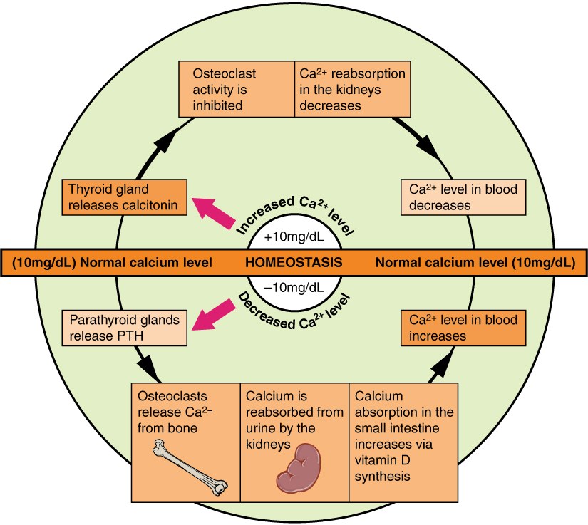
Hormones set the background rate of remodeling and control blood calcium.
| Hormone | Main effect on bone | Clinical point |
|---|---|---|
| Parathyroid hormone (PTH) | Acts on osteoblasts and osteocytes; raises RANKL; releases calcium | Constantly high PTH removes bone. Short daily pulses increase formation: teriparatide reduced vertebral fractures by about two-thirds (Neer 2001). |
| Vitamin D | Increases absorption of calcium and phosphate from the gut | Deficiency causes poor mineralization (rickets, osteomalacia) |
| Calcitonin | Directly inhibits osteoclasts | Small role in adults |
| Estrogen | Restrains osteoclasts; keeps bone cells alive | Loss at the menopause speeds bone loss |
| Androgens | Increase periosteal bone formation; part of their effect is through conversion to estrogen | Men have wider bones |
| Growth hormone and IGF-1 | Increase formation and turnover | Needed for growth |
| Thyroid hormone | Increases turnover | Excess causes net bone loss |
| Glucocorticoids in excess | Reduce osteoblast formation and survival; kill osteocytes | The commonest cause of secondary osteoporosis; also slows fracture healing |
| FGF23 | Increases phosphate loss in the urine | Excess causes rickets and osteomalacia |
6.7 Drugs that change remodeling
Many older patients, and many patients with cancer, take drugs that act on remodeling. The reconstructive surgeon must ask about them, because they change how the jaw heals.
| Drug | Action | Main use |
|---|---|---|
| Bisphosphonates (alendronate, zoledronic acid) | Bind to bone mineral and are taken up by osteoclasts, which then die. They stay in bone for years. | Osteoporosis; bone metastases; myeloma |
| Denosumab | Antibody against RANKL; stops osteoclast formation. Reduced vertebral fractures by 68% over three years (Cummings 2009). The effect wears off within months of stopping. | Osteoporosis; bone metastases |
| Teriparatide, abaloparatide | Daily pulses of PTH action; increase formation | Severe osteoporosis |
| Romosozumab | Antibody against sclerostin; increases formation and reduces removal. Reduced new vertebral fractures by 73% at one year (Cosman 2016). | Severe osteoporosis |
6.7.1 Medication-related osteonecrosis of the jaw
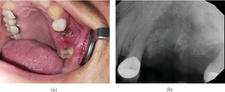
The jaws remodel quickly and are covered only by thin mucosa. When remodeling is strongly suppressed, an extraction socket or an ulcer may not heal, and bone becomes exposed. This is medication-related osteonecrosis of the jaw (MRONJ). It is defined as exposed bone in the jaw, or bone that can be probed through a fistula, lasting more than 8 weeks, in a patient who has taken anti-resorptive or anti-angiogenic drugs and has had no radiotherapy to the jaws (Ruggiero 2022).
- The risk is much higher with the high doses used for cancer (for example monthly zoledronic acid or denosumab) than with the doses used for osteoporosis.
- Extraction of teeth is the commonest trigger.
- Advanced disease is treated by resecting the dead bone. In some patients this needs reconstruction with a vascularized bone flap.
- Before elective jaw surgery, dental implants or a fibula flap, ask whether the patient has taken these drugs, which drug, at what dose and for how long.
Osteoradionecrosis and MRONJ look similar but have different causes. In osteoradionecrosis the bone has lost its vessels and cells. In MRONJ the vessels may be present, but remodeling and repair are suppressed.
6.8 Abnormal states of bone
| State | The basic problem | How to tell it apart |
|---|---|---|
| Osteoporosis | Too little bone, with damaged structure | The remaining bone is normally mineralized |
| Osteomalacia | Osteoid not mineralized | Wide osteoid seams; low vitamin D or phosphate |
| Osteopetrosis | Osteoclasts do not work | Very dense but brittle bone; small marrow space; jaw osteomyelitis |
| Disuse bone loss | Loss of load | Follows immobilization, paralysis or stress shielding |
| Hyperparathyroidism, hyperthyroidism | High turnover with net loss | Formation markers are high but bone is still lost |
6.9 Summary
- Modeling changes the shape of bone; remodeling renews it in place; repair restores continuity after injury.
- The remodeling cycle is activation, resorption (weeks), reversal, formation (months) and mineralization.
- Because removal is fast and formation slow, more remodeling means weaker bone for a time. Net gain or loss depends on balance at each site.
- Osteocytes sense load through fluid flow. Load reduces sclerostin and lets bone form; disuse raises sclerostin and RANKL.
- Bone keeps only what is loaded: stress shielding under plates, hypertrophy of a loaded fibula, resorption of unloaded grafts and of the edentulous alveolus.
- PTH, vitamin D, estrogen and glucocorticoids set the rate of remodeling. Intermittent PTH builds bone.
- Anti-resorptive drugs can cause osteonecrosis of the jaw. Ask about them before jaw surgery.
References
- Hauge EM, Qvesel D, Eriksen EF, Mosekilde L, Melsen F. Cancellous bone remodeling occurs in specialized compartments lined by cells expressing osteoblastic markers. J Bone Miner Res 2001;16:1575–1582.
- Parfitt AM. Osteonal and hemi-osteonal remodeling: the spatial and temporal framework for signal traffic in adult human bone. J Cell Biochem 1994;55:273–286.
- Robling AG, Niziolek PJ, Baldridge LA, Condon KW, Allen MR, Alam I, et al. Mechanical stimulation of bone in vivo reduces osteocyte expression of Sost/sclerostin. J Biol Chem 2008;283:5866–5875.
- Wang L, You X, Lotinun S, Zhang L, Wu N, Zou W. Mechanical sensing protein PIEZO1 regulates bone homeostasis via osteoblast-osteoclast crosstalk. Nat Commun 2020;11:282.
- Frost HM. Bone's mechanostat: a 2003 update. Anat Rec A Discov Mol Cell Evol Biol 2003;275:1081–1101.
- Burr DB, Martin RB, Schaffler MB, Radin EL. Bone remodeling in response to in vivo fatigue microdamage. J Biomech 1985;18:189–200.
- Uhthoff HK, Dubuc FL. Bone structure changes in the dog under rigid internal fixation. Clin Orthop Relat Res 1971;81:165–170.
- Neer RM, Arnaud CD, Zanchetta JR, Prince R, Gaich GA, Reginster JY, et al. Effect of parathyroid hormone (1-34) on fractures and bone mineral density in postmenopausal women with osteoporosis. N Engl J Med 2001;344:1434–1441.
- Cummings SR, San Martin J, McClung MR, Siris ES, Eastell R, Reid IR, et al. Denosumab for prevention of fractures in postmenopausal women with osteoporosis. N Engl J Med 2009;361:756–765.
- Cosman F, Crittenden DB, Adachi JD, Binkley N, Czerwinski E, Ferrari S, et al. Romosozumab treatment in postmenopausal women with osteoporosis. N Engl J Med 2016;375:1532–1543.
- Ruggiero SL, Dodson TB, Aghaloo T, Carlson ER, Ward BB, Kademani D. American Association of Oral and Maxillofacial Surgeons' position paper on medication-related osteonecrosis of the jaws: 2022 update. J Oral Maxillofac Surg 2022;80:920–943.