13.1 Introduction and definition
Distraction osteogenesis makes new bone grow at the site of the deficiency, without a graft.
Definition: distraction osteogenesis is the formation of new bone between two bone segments that are slowly pulled apart after an osteotomy.
The steps are: cut the bone, wait a few days for early callus to form, then pull the two segments apart slowly (about 1 mm per day) with a device. The callus is stretched and keeps forming new bone in the gap.
The main advantage for plastic surgery: the soft tissues (skin, muscle, nerves, vessels, mucosa) are stretched and grow together with the bone. With a bone graft, the soft tissue is often too tight to cover the added bone. With distraction, the soft tissue is expanded at the same time.
Flowchart: Overview of distraction osteogenesis
- Osteotomy
- Latency5 to 7 days; wait for callus
- Distraction1 mm per day
- Consolidation6 to 8 weeks or more
- Device removed
13.2 History
- 1905, Codivilla: first lengthening of the femur by traction after osteotomy.
- 1950s onward, Ilizarov: developed the method in limbs and described the "tension-stress effect": slow, steady traction on living tissue stimulates it to grow (Ilizarov 1989).
- 1973, Snyder: lengthened the mandible in dogs.
- 1992, McCarthy: first mandibular distraction in humans: 4 children, lengthened by 18 to 24 mm.
- After 1995: extended to the midface, alveolus and cranial vault.
13.3 The three phases
| Phase | What is done | What happens in the bone | Usual duration |
|---|---|---|---|
| 1. Latency | Wait after the osteotomy; device not turned | Hematoma and early soft callus form | 5 to 7 days in adults; 0 to 2 days in neonates |
| 2. Distraction (activation) | Device turned every day | Callus is stretched; new bone forms in the gap | About 1 mm per day until the planned length is reached |
| 3. Consolidation | Device left in place, not turned | New bone mineralizes and becomes strong | At least twice the distraction period; usually 6 to 8 weeks or more |
Then the device is removed.
13.4 Rate and rhythm
The three phases are controlled by three settings: the latency period, the rate (millimeters per day) and the rhythm (how many times per day the device is turned).
Ilizarov tested different rates in dogs (Ilizarov 1989):
- 0.5 mm per day: too slow. The bone united before the lengthening was finished (premature consolidation).
- 1 mm per day: best result.
- 2 mm per day: too fast. Bone formation could not keep up, and the gap filled with fibrous tissue.
- Dividing the daily amount into several small steps (for example 0.25 mm four times a day) gave better bone than one large step.
Flowchart: Effect of distraction rate
- Rate of distraction
- Too slow0.5 mm per day
- Premature consolidationbone unites before target length
- Correct1 mm per day, in several small steps
- Good bone regenerate
- Too fast2 mm per day
- Fibrous tissue in the gapnonunion
- Too slow0.5 mm per day
Children heal faster than adults. In neonates, latency is short or omitted and the rate may be 1 to 2 mm per day, to avoid premature consolidation.
13.5 Histology of the distraction gap
The rate must match the speed of bone formation. This section describes the tissue that forms in the gap.
Figure 13.2. Zones of the distraction gap (the regenerate). The zones are the same on both sides of the center.
- Fibrous interzone (center): dividing cells and collagen fibers, lined up in the direction of distraction. This is where new tissue is produced.
- Mineralization zone: bone forms directly on the collagen fibers, as columns that grow toward the center.
- Remodeling zone (next to the host bone): the new bone matures.
Bone forms mainly by intramembranous ossification (directly, without cartilage), provided the device is stable (Karp 1992). If the fixation is unstable, cartilage and fibrous tissue form instead.
13.5.1 Biology of the regenerate
- Mechanotransduction: tension on the cells of the interzone activates integrin and stretch-sensitive pathways, which increase cell proliferation and matrix production.
- Growth factors are increased throughout distraction: BMP-2 and BMP-4, TGF-β1, FGF-2, IGF-1 and VEGF.
- Blood flow in the distracted bone rises to several times normal during distraction and stays raised during consolidation (Aronson 1994). Angiogenesis is essential: the new vessels grow parallel to the bone columns.
- Distraction histogenesis: skin, muscle, nerves and vessels also grow under tension. Muscle is the tissue that limits the rate, because it adapts most slowly.
- Corticotomy (cutting the cortex and preserving the marrow and periosteum) was Ilizarov's original technique. A complete osteotomy with careful preservation of periosteum gives similar results.
13.5.2 Devices
| Type | Advantages | Disadvantages |
|---|---|---|
| External | Long distraction possible; vector can be adjusted during treatment (multiplanar devices); easy removal | Facial scars from pin tracks; pin loosening; visible; easily knocked |
| Internal (buried) | No visible device except the activation arm; better tolerated; more stable | Second operation for removal; fixed vector; limited length |
| Resorbable or semi-buried | Avoids a second operation | Less rigid; limited availability |
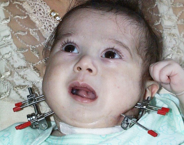
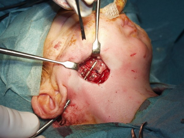
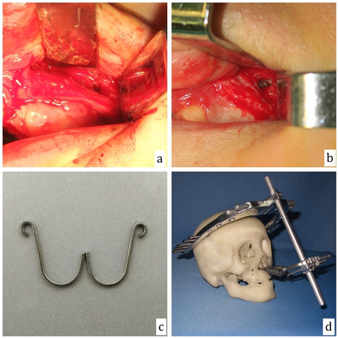
13.6 The vector
The bone lengthens in the direction of the device. This direction is called the vector. It must be planned before surgery, because it is decided by where the device is fixed.
| Vector | Lengthens | Example |
|---|---|---|
| Vertical | Ramus | Hemifacial microsomia |
| Horizontal | Body | Pierre Robin sequence |
| Oblique | Both | Combined deficiency |
In children, the osteotomy and the screws must avoid the tooth buds and the inferior alveolar nerve.
Figure 13.3. Vectors of mandibular distraction.
13.7 Uses in plastic and craniofacial surgery
Flowchart: Uses of distraction in plastic surgery
- Distraction osteogenesis
- Mandible
- Pierre Robin sequence
- Hemifacial microsomia
- Maxilla and midface
- Cleft maxillary hypoplasiaLe Fort I
- Syndromic craniosynostosisLe Fort III, monobloc
- Cranial vault
- Posterior vault expansion
- Alveolus and hand
- Ridge augmentation; thumb lengthening
- Mandible
13.7.1 Mandible: Pierre Robin sequence
In Pierre Robin sequence the mandible is small, the tongue falls back and blocks the airway. In severe cases the traditional treatment was tracheostomy. Mandibular distraction lengthens the mandible and moves the tongue base forward, because the tongue muscles are attached to the mandible.
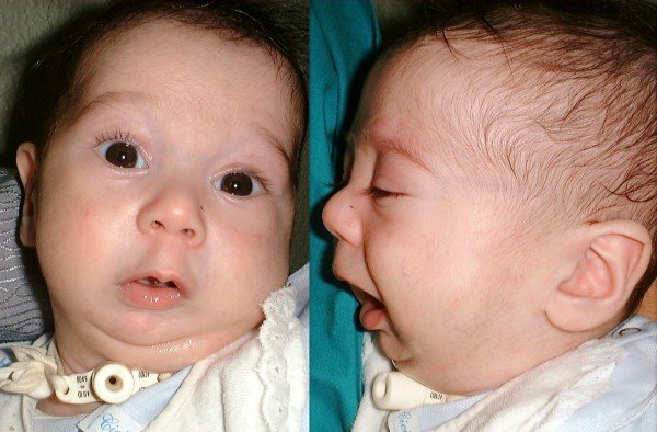
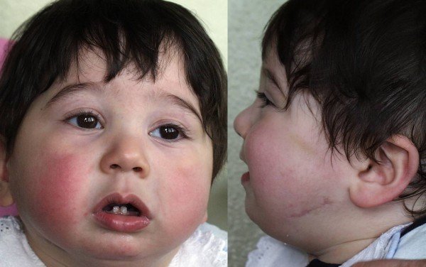
Figure 13.4. Left: micrognathia in Pierre Robin sequence, with tracheostomy. Right: another infant after mandibular distraction. Ramieri et al., Ital J Pediatr 2012, CC BY 2.0.
- Denny (2001): 10 young children with airway obstruction. After distraction of 8 to 22 mm, the airway area increased by a mean of 67%. All improved clinically.
- Greathouse (2016): compared distraction (74 infants) with tongue-lip adhesion (15 infants). Success was 90.5% with distraction and 60% with tongue-lip adhesion.
- Breik (2016), systematic review: after distraction, 82% of children fed completely by mouth.
Selection for mandibular distraction in Pierre Robin sequence:
- Most infants are managed without surgery: prone positioning, nasopharyngeal airway.
- Surgery is considered when there is persistent obstruction (desaturation, raised carbon dioxide, obstructive events on polysomnography) or failure to feed and gain weight.
- Airway endoscopy is done first. Distraction treats obstruction at the tongue base only. It does not treat laryngomalacia, tracheomalacia or central apnea, and in those cases tracheostomy may still be needed.
- Typical neonatal protocol: latency 0 to 2 days, rate 1 to 2 mm per day, until the lower alveolus is slightly ahead of the upper (overcorrection), consolidation 4 to 8 weeks.
Hemifacial microsomia (Pruzansky classification, modified by Kaban):
| Type | Mandible and TMJ | Usual treatment |
|---|---|---|
| I | Small but normally shaped ramus and condyle | Observation, orthodontics; orthognathic surgery at maturity |
| IIA | Abnormal shape of ramus and condyle, but the joint is in an acceptable position and functions | Distraction of the ramus, or orthognathic surgery |
| IIB | Ramus and condyle hypoplastic and displaced; no functioning joint | Reconstruction of ramus and condyle with a costochondral graft (or vascularized bone) |
| III | Ramus, condyle and glenoid fossa absent | Costochondral graft and fossa reconstruction; later distraction of the graft or orthognathic surgery |
The long-term stability of early distraction in hemifacial microsomia is debated. Asymmetry tends to return with growth, and many patients still need surgery at skeletal maturity.
13.7.2 Other sites
| Site | Indication | Why distraction is used |
|---|---|---|
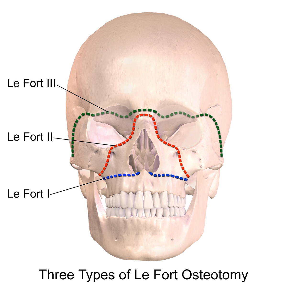
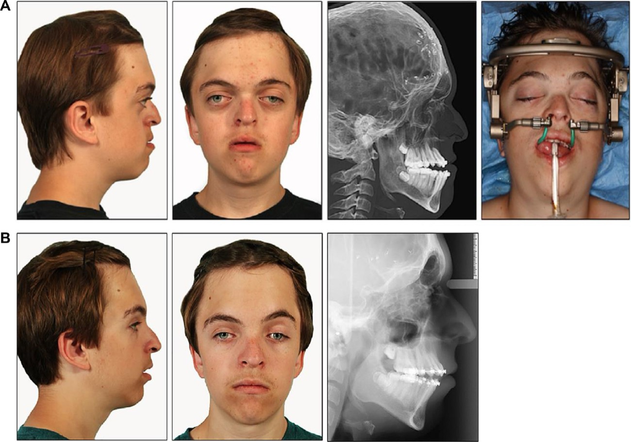
| Maxilla (Le Fort I) | Severe maxillary hypoplasia in cleft patients | The scarred palate limits a large one-stage advancement and causes relapse (Polley and Figueroa 1997; Cheung and Chua 2006) |
| Midface (Le Fort III, monobloc) | Syndromic craniosynostosis (Crouzon, Apert) | Large advancement; less dead space and infection than one-stage monobloc (Arnaud 2007: 36 children) |
| Cranial vault | Posterior vault expansion in craniosynostosis | The scalp expands with the bone, allowing more volume (White 2009) |
| Alveolus | Deficient alveolar ridge before implants | Gains bone and gingiva together (Chin and Toth 1996) |
| Hand | Thumb or metacarpal lengthening after amputation | No donor site (Matev 1989) |
13.8 Complications
| Phase | Complication | Cause |
|---|---|---|
| Operation | Injury to tooth buds or inferior alveolar nerve; incomplete osteotomy | Crowded bone in children |
| Distraction | Pin-site infection; device loosening or failure; wrong vector; premature consolidation; fibrous nonunion | Thin bone; wrong device position; rate too slow or too fast |
| Consolidation and after | Weak regenerate, relapse, open bite, scars | Device removed too early |
13.9 Summary
- Distraction osteogenesis forms new bone by slowly separating two bone segments after osteotomy.
- Three phases: latency, distraction, consolidation.
- Standard rate is 1 mm per day. Too fast gives fibrous nonunion. Too slow gives premature consolidation.
- The soft tissue expands with the bone. This is why it is so useful in craniofacial surgery.
References
- Denny AD, Talisman R, Hanson PR, Recinos RF. Mandibular distraction osteogenesis in very young patients to correct airway obstruction. Plast Reconstr Surg 2001;108:302–311.
- Codivilla A. On the means of lengthening, in the lower limbs, the muscles and tissues which are shortened through deformity. Am J Orthop Surg 1905;2:353–369.
- Ilizarov GA. The tension-stress effect on the genesis and growth of tissues. Part I. The influence of stability of fixation and soft-tissue preservation. Clin Orthop Relat Res 1989;238:249–281.
- Ilizarov GA. The tension-stress effect on the genesis and growth of tissues. Part II. The influence of the rate and frequency of distraction. Clin Orthop Relat Res 1989;239:263–285.
- Snyder CC, Levine GA, Swanson HM, Browne EZ Jr. Mandibular lengthening by gradual distraction: preliminary report. Plast Reconstr Surg 1973;51:506–508.
- McCarthy JG, Schreiber J, Karp N, Thorne CH, Grayson BH. Lengthening the human mandible by gradual distraction. Plast Reconstr Surg 1992;89:1–8.
- Karp NS, McCarthy JG, Schreiber JS, Sissons HA, et al. Membranous bone lengthening: a serial histological study. Ann Plast Surg 1992;29:2–7.
- McCarthy JG, Stelnicki EJ, Mehrara BJ, Longaker MT. Distraction osteogenesis of the craniofacial skeleton. Plast Reconstr Surg 2001;107:1812–1827.
- Molina F, Ortiz Monasterio F. Mandibular elongation and remodeling by distraction: a farewell to major osteotomies. Plast Reconstr Surg 1995;96:825–840.
- Polley JW, Figueroa AA. Management of severe maxillary deficiency in childhood and adolescence through distraction osteogenesis with an external, adjustable, rigid distraction device. J Craniofac Surg 1997;8:181–185.
- Cheung LK, Chua HDP. A meta-analysis of cleft maxillary osteotomy and distraction osteogenesis. Int J Oral Maxillofac Surg 2006;35:14–24.
- Arnaud E, Marchac D, Renier D. Reduction of morbidity of the frontofacial monobloc advancement in children by the use of internal distraction. Plast Reconstr Surg 2007;120:1009–1026.
- White N, Evans M, Dover MS, Noons P, Solanki G, Nishikawa H. Posterior calvarial vault expansion using distraction osteogenesis. Childs Nerv Syst 2009;25:231–236.
- Chin M, Toth BA. Distraction osteogenesis in maxillofacial surgery using internal devices: review of five cases. J Oral Maxillofac Surg 1996;54:45–53.
- Matev IB. The bone-lengthening method in hand reconstruction: twenty years' experience. J Hand Surg Am 1989;14:376–378.
- Greathouse ST, Costa M, Ferrera A, et al. The surgical treatment of Robin sequence. Ann Plast Surg 2016;77:413–419.
- Breik O, Umapathysivam K, Tivey D, Anderson P. Feeding and reflux in children after mandibular distraction osteogenesis for micrognathia: a systematic review. Int J Pediatr Otorhinolaryngol 2016;85:128–135.
- Aronson J. Temporal and spatial increases in blood flow during distraction osteogenesis. Clin Orthop Relat Res 1994;301:124–131.