4.1 Introduction
Bone is a very vascular tissue. Its cells need blood within a short distance, and they die within hours to days when the supply stops. New bone cannot form without new vessels.
For the reconstructive surgeon, the blood supply of bone is the basis of three daily decisions: how much periosteum to strip, whether a bone fragment or graft will survive, and when to choose a vascularized bone flap instead of a graft.
Flowchart: Overview of the blood supply of bone
- Blood supply of bone
- From inside
- Nutrient arterymarrow and inner cortex
- Metaphyseal and epiphyseal arteriesthe ends of the bone
- From outside
- Periosteal vesselsouter cortex
- Vessels of attached muscle, fascia, mucosa and duramain supply of flat and facial bones
- Linked by vessels that cross the cortex
- From inside
4.2 Arterial supply of a long bone
A long bone has three main arterial systems (Trueta and Morgan 1960; Rhinelander 1974):
| System | Course | What it supplies |
|---|---|---|
| Nutrient (medullary) artery | Enters the shaft through the nutrient foramen, then divides into ascending and descending branches in the marrow | The marrow and the inner two-thirds of the cortex |
| Metaphyseal and epiphyseal arteries | Many small vessels from the arteries around the joint, entering near the ends of the bone | The metaphysis and epiphysis. In the adult they join the nutrient system. |
| Periosteal arteries | A network on the bone surface, fed mainly where muscles and fascia attach | The outer third of the cortex |
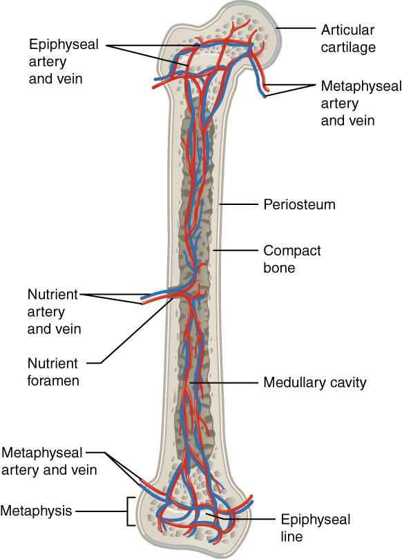
Three points about this pattern matter in surgery:
- Flow in the cortex normally runs from inside to outside, from the marrow toward the periosteum.
- The systems are connected. When the nutrient artery is destroyed, for example by a fracture or by reaming for a nail, the periosteal vessels take over more of the cortex and flow can reverse (Rhinelander 1974). A fracture that has lost its nutrient supply then depends on the periosteum and the surrounding muscle.
- The "inner two-thirds and outer one-third" rule is only a guide. It does not mean the outer cortex or the periosteum can be stripped freely when the nutrient artery is intact.
The position of the nutrient foramen varies between bones and between people. In the fibula it is usually near the middle of the shaft. This is why the middle of the fibula is chosen for a flap that will be cut into segments: the segment containing the nutrient artery gets both supplies, and the others survive on the periosteal branches.
4.3 Vessels inside the cortex
Inside the cortex, blood runs in the central (Haversian) canals along the bone and in the perforating (Volkmann) canals across it. The osteocytes are fed from these vessels through the fluid in their canaliculi.
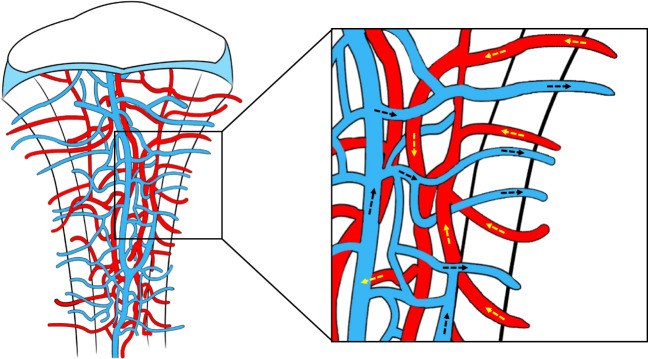
Recent imaging has shown that the cortex of a long bone is crossed by a large number of small vessels running straight from the marrow to the periosteum. In mice, these trans-cortical vessels carry more than 80% of arterial blood and 59% of venous blood leaving the bone, and similar vessels were found in human limb bones (Grüneboom 2019). The inner and outer systems are therefore one connected network, not two separate ones.
4.4 Periosteal and soft tissue supply
The periosteum has its own network of vessels. It is fed by the muscles, fascia and ligaments attached to it. Branches pass from it into the outer cortex. This means:
- A bone fragment that remains attached to muscle or periosteum can stay alive even when it has no medullary supply.
- Stripping periosteum removes this supply, and stripping the muscle off the periosteum removes its feeding vessels.
- In a bone flap, the periosteum and a cuff of muscle are kept on the bone because they carry the vessels.
The fibula is the clearest example. Its nutrient artery and its periosteal branches both come from the peroneal artery. The periosteal branches enter at intervals along the bone through the muscles and the intermuscular septum. When the fibula is cut into segments for a mandible, each segment survives because it keeps its periosteal branches from the peroneal artery, even though only one segment contains the nutrient artery.
4.5 Venous drainage
Blood leaves the marrow through large thin-walled channels (sinusoids) that drain into veins running with the nutrient artery, into veins at the ends of the bone, and into periosteal veins. In the skull, the diploic veins drain the diploë.
A bone flap needs venous outflow as much as arterial inflow. In free flaps, venous thrombosis is more common than arterial thrombosis. In the fibula flap, the two venae comitantes of the peroneal artery drain the bone.
4.6 Blood supply of flat and facial bones
Flat and facial bones are supplied differently from long bones in one important way: most of their blood comes from the soft tissue attached to them, and little or none comes from a nutrient artery.
- A flat bone has no marrow cavity and usually no large nutrient artery.
- Many small vessels enter from the periosteum and from the muscles, mucosa or dura attached to it.
- Inside the bone, these vessels join in the cancellous layer.
Flowchart: Long bone compared with flat and facial bone
- Main blood supply
- Long bone
- Mainly from insidenutrient artery to marrow and inner cortex; periosteum to outer cortex
- Flat and facial bone
- Mainly from attached soft tissueperiosteum, muscle, mucosa and dura
- The bone stays alive as long as its soft tissue stays attached
- Mainly from attached soft tissueperiosteum, muscle, mucosa and dura
- Long bone
| Bone | Blood supply | What it means for the surgeon |
|---|---|---|
| Calvarium | Inner table: from the dura, mainly the middle meningeal artery. Outer table: from the pericranium and scalp, through the superficial temporal, occipital and supraorbital arteries. Veins drain through the diploë. | A piece of skull lifted off both the dura and the pericranium is a free graft. If it is left attached to the pericranium or temporoparietal fascia with the superficial temporal vessels, it is a vascularized calvarial flap (McCarthy and Zide 1984). |
| Mandible | Inside: the inferior alveolar artery in the canal. Outside: the periosteum and the attached muscles (masseter, medial pterygoid, floor of mouth), fed by the facial, lingual and maxillary arteries. | In the young dentate mandible the inferior alveolar artery is the main supply. With age and loss of teeth it narrows, and the mandible depends mainly on its periosteum (Bradley 1972). In the atrophic edentulous mandible, strip as little periosteum as possible. |
| Maxilla | No single nutrient artery. Many vessels reach it through the mucosa and periosteum: posterior superior alveolar, infraorbital, descending palatine and nasopalatine arteries, and the ascending palatine and ascending pharyngeal arteries through the soft palate. | After a Le Fort I osteotomy the descending palatine arteries are often cut, and the maxilla survives on its palatal and buccal soft tissue (Bell 1975). The risk of ischemia is higher when the maxilla is cut into several pieces, and in a cleft maxilla with palatal scar. |
| Zygoma, nasal bones, orbital walls | Small periosteal vessels from the surrounding soft tissue | Facial fracture fragments usually stay alive because they stay attached to periosteum. Do not discard fragments that still have a soft tissue attachment. |
| Ilium, scapula, rib | Periosteal and muscular vessels from a named artery: deep circumflex iliac artery for the iliac crest, circumflex scapular artery for the lateral border of the scapula, posterior intercostal artery for the rib | These bones can be moved as flaps if their periosteum and a cuff of muscle are kept on them. |
This rich supply from the soft tissue is one reason why facial bones heal faster than long bones and are less often infected. It is lost after radiotherapy and in scarred tissue, and in those conditions facial bone heals poorly.
4.7 Blood supply of growing bone
The physis (growth plate) is cartilage and has no vessels of its own. It depends on the vessels on either side:
- Epiphyseal vessels feed the resting and proliferative zones from the epiphyseal side. If they are damaged, the cartilage cells die and growth stops.
- Metaphyseal vessels end in loops at the bottom of the physis. They invade the calcified cartilage and bring the cells that replace it with bone.
This is why a vascularized fibular epiphyseal transfer in a child must be based on the vessel that supplies the proximal fibular epiphysis (the anterior tibial artery). Without that vessel, the physis is carried but does not grow.
The slow flow in the metaphyseal loops of children is also why blood-borne bone infection in children usually starts in the metaphysis.
4.8 New vessels and new bone
New bone forms only where new vessels have grown. This link is called angiogenic-osteogenic coupling.
Flowchart: How new vessels lead to new bone
- Low oxygen (hypoxia)after a fracture, or in growing cartilage
- HIF-1α becomes activethe cell's oxygen sensor
- VEGF is releasedvessel growth factor
- New capillaries grow inbringing osteoprogenitors on their surface
- New bone forms
In mice, a specific type of capillary, called type H, lies next to growing bone and carries the progenitor cells that form it. These vessels decline with age, and restoring them restored bone mass in old mice (Kusumbe 2014; Ramasamy 2014). This is laboratory work; it explains why vessels and bone grow together, but it is not yet a treatment.
Brief hypoxia starts this process. Prolonged ischemia stops it. A fracture with poor blood supply or an irradiated bed produces fibrous tissue or cartilage instead of bone.
4.9 When the blood supply fails
4.9.1 The recipient bed
A free (non-vascularized) bone graft has no blood supply when it is placed. Most of its cells die. It depends on the vessels of the recipient bed to grow into it and bring new cells. The quality of the bed therefore decides whether a free graft survives.
| Recipient bed | Problem | Usual answer |
|---|---|---|
| Healthy, well vascularized | None | A free graft can incorporate |
| Scarred | Few vessels | Excise scar; bring in vascularized tissue |
| Irradiated | Few vessels, few cells, low oxygen | Vascularized bone flap, or graft covered by a flap |
| Infected | Dead tissue, inflammation | Debride first; then reconstruct, often with a flap |
| Poor soft tissue cover | Graft exposed | Soft tissue cover is part of the bone reconstruction |
4.9.2 Radiotherapy and osteoradionecrosis
Radiotherapy damages the small vessels and the cells of bone. Marx (1983) described the irradiated bone as hypoxic, hypocellular and hypovascular. In his series, bacteria were only contaminants; the primary problem was tissue that could not repair itself. Minor injury, such as a dental extraction, can then lead to a wound that does not heal and to exposed dead bone: osteoradionecrosis.
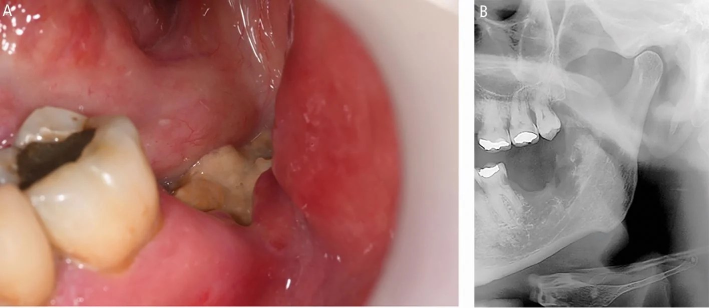
This is the main reason why the head and neck reconstructive surgeon uses vascularized bone. A free fibula or iliac crest flap brings its own blood supply into a bed that cannot supply a graft. Advanced osteoradionecrosis of the mandible is treated by resecting the dead bone and replacing it with a vascularized bone flap.
4.10 Vascularized bone flaps
A vascularized bone flap keeps the bone's own vessels, which are joined to vessels at the recipient site. The osteocytes stay alive, so the flap heals to the host bone like a fracture and does not depend on the bed. Each flap is supplied in a different way.
| Flap | Artery | How the bone is supplied |
|---|---|---|
| Fibula | Peroneal | Nutrient artery and segmental periosteal branches |
| Iliac crest | Deep circumflex iliac | Branches through the inner (iliacus) muscle cuff and periosteum of the inner table |
| Scapula | Circumflex scapular; angular branch of the thoracodorsal | Periosteal branches to the lateral border and the tip |
| Radius | Radial | Small periosteal branches through the intermuscular septum |
| Medial femoral condyle | Descending genicular | Periosteal network on the condyle |
| Calvarium | Superficial temporal | Through the temporoparietal fascia and pericranium |
A vascularized flap still needs good fixation, contact with the host bone and good soft tissue closure. Its vessels must stay open: both the artery and the veins.
4.11 Summary
- A long bone is supplied by the nutrient artery, metaphyseal and epiphyseal arteries, and periosteal vessels. Normal flow in the cortex runs from inside to outside.
- The systems are connected by many vessels crossing the cortex. When one is lost, the others take over part of its territory.
- Flat and facial bones depend mainly on the soft tissue attached to them. Keep facial bone attached to its periosteum.
- The mandible relies more on its periosteum with age and loss of teeth. The maxilla survives a Le Fort I osteotomy on its palatal and buccal soft tissue.
- The physis depends on epiphyseal vessels for growth and metaphyseal vessels for ossification.
- New bone forms only where new vessels grow (hypoxia, HIF-1α, VEGF).
- Irradiated bone is hypoxic, hypocellular and hypovascular. It needs vascularized tissue.
References
- Trueta J, Morgan JD. The vascular contribution to osteogenesis. I. Studies by the injection method. J Bone Joint Surg Br 1960;42-B:97–109.
- Rhinelander FW. Tibial blood supply in relation to fracture healing. Clin Orthop Relat Res 1974;(105):34–81.
- Grüneboom A, Hawwari I, Weidner D, Culemann S, Müller S, Henneberg S, et al. A network of trans-cortical capillaries as mainstay for blood circulation in long bones. Nat Metab 2019;1:236–250.
- Chen M, Li Y, Huang X, Gu Y, Li S, Yin P, et al. Skeleton-vasculature chain reaction: a novel insight into the mystery of homeostasis. Bone Res 2021;9:21.
- McCarthy JG, Zide BM. The spectrum of calvarial bone grafting: introduction of the vascularized calvarial bone flap. Plast Reconstr Surg 1984;74:10–18.
- Bradley JC. Age changes in the vascular supply of the mandible. Br Dent J 1972;132:142–144.
- Bell WH, Fonseca RJ, Kennedy JW, Levy BM. Bone healing and revascularization after total maxillary osteotomy. J Oral Surg 1975;33:253–260.
- Kusumbe AP, Ramasamy SK, Adams RH. Coupling of angiogenesis and osteogenesis by a specific vessel subtype in bone. Nature 2014;507:323–328.
- Ramasamy SK, Kusumbe AP, Wang L, Adams RH. Endothelial Notch activity promotes angiogenesis and osteogenesis in bone. Nature 2014;507:376–380.
- Marx RE. Osteoradionecrosis: a new concept of its pathophysiology. J Oral Maxillofac Surg 1983;41:283–288.