8B.1 Introduction
Orthobiologics are biological substances used to help bone and other musculoskeletal tissues heal. They are made from the patient's own blood or marrow, or produced by recombinant technology.
A bone graft supplies several things at once. An orthobiologic usually supplies one of them: a signal, or cells, or a vascular bed. For this reason an orthobiologic is almost always an addition to a graft and to good surgery. It does not replace them.
Two points apply to the whole chapter:
- Most of the clinical evidence comes from orthopedic and spinal surgery. Evidence in plastic and craniofacial surgery is limited, and is stated where it exists.
- Many of these products were promoted before the evidence was complete. Each section therefore states what has been shown and what has not.
Flowchart: Overview of orthobiologics
- Orthobiologics
- Supply a signal
- Bone morphogenetic proteinrhBMP-2, rhBMP-7
- Platelet concentratesPRP, PRF
- Systemic drugsteriparatide
- Supply cells
- Bone marrow aspirate and concentrate
- Adipose-derived stem cells
- Combine all of these
- Tissue engineeringscaffold, cells and signal
- Create a vascular bed
- Induced membrane techniqueMasquelet
- Supply a signal
8B.1.1 Where they fit, in brief
The table gives the verdict on each product before the details. The flowchart shows what must be in place before any of them is considered.
| Product | Supplies | Does not supply | Strength of evidence for bone | Use in plastic surgery |
|---|---|---|---|---|
| rhBMP-2 | Osteoinductive signal | Cells, scaffold strength, blood supply | Randomized trials in tibia, spine and sinus; small series in cleft and mandible | Selected small jaw defects in adults; off-label; not near tumor, not in young children |
| PRP, PRF | Supportive growth factors; a fibrin membrane | Osteoinduction, cells, scaffold | Weak and inconsistent | Optional addition to a graft; soft tissue healing |
| Marrow aspirate or concentrate | Cells, some signal | Scaffold, strength | Case series; dose-dependent | Added to allograft or ceramic in small defects |
| Adipose-derived stem cells | Cells | Scaffold, strength | Case reports and small series | Experimental |
| Induced membrane | A vascular bed that releases growth factors | Bone: autograft is still needed | Many series; about one in five need further surgery | Flap cover in stage 1; whole technique in the hand |
| Teriparatide | Systemic stimulus to osteoblasts | Anything local | One trial with an unclear result | None established |
Flowchart: Before using an orthobiologic
- Is the defect stable?if not, fix it
- Is the bed vascular and covered?if not, bring a flap
- Is the defect small enough for a graft?if not, vascularized bone
- Only then: would a signal or cells add something?
Where possible, each product is described in the same order: what it is, how it works, examples of products, evidence, risks, and its position in plastic surgery.
8B.2 Products that supply a signal
8B.2.1 Growth factors in bone healing
A growth factor is a protein that binds to a receptor on a cell and changes what the cell does. Several are active during bone healing. Each has a different role.
| Growth factor | Source | Main action on bone |
|---|---|---|
| BMP (bone morphogenetic proteins) | Bone matrix, osteoprogenitors | Osteoinduction: makes a mesenchymal stem cell (MSC) become an osteoblast. The only family that can induce bone in a non-bone site. |
| TGF-β | Platelets, bone matrix | MSC proliferation, matrix production; cartilage formation |
| PDGF | Platelets, macrophages | Attracts MSCs and osteoblasts and makes them divide |
| FGF (FGF-2) | Macrophages, MSCs | New vessels; proliferation of progenitor cells |
| IGF-1 | Bone matrix, liver | Osteoblast proliferation and matrix synthesis |
| VEGF | Hypertrophic chondrocytes, osteoblasts (through HIF-1α) | New vessels; links vessel growth and bone formation |
The distinction that matters clinically:
Flowchart: Two kinds of signal
- Growth factors in bone healing
- Osteoinductive
- BMPcan start bone formation, even outside bone
- Supportive (not osteoinductive)
- PDGF, TGF-β, FGF, IGF, VEGFrecruit cells, increase their number and bring vessels, where bone is already forming
- Osteoinductive
BMP products supply the first kind. Platelet products supply the second kind. This explains much of the difference in their results.
8B.2.2 Bone morphogenetic protein (BMP)
What it is
BMPs are growth factors of the TGF-β family. They make stem cells become osteoblasts. They were identified from Urist's observation that demineralized bone placed in muscle forms new bone.
How it works
- More than 20 BMPs are known. BMP-2, 4, 6, 7 and 9 are the most osteogenic. BMP-3 inhibits bone formation. BMP-1 is not a true BMP; it is a protease.
- Related pathway: Wnt/β-catenin also drives osteoblast differentiation. It is inhibited by sclerostin (from osteocytes) and DKK-1.
- Natural inhibitors: noggin, chordin and gremlin outside the cell; Smad 6 and 7 inside the cell.
Flowchart: BMP signaling
- BMP binds its receptorstype I and type II serine/threonine kinase receptors
- Smad 1, 5 and 8 are phosphorylated
- They join Smad 4 and enter the nucleus
- Runx2 and Osterix are switched onmaster genes of the osteoblast
- The stem cell becomes an osteoblast
Products and how they are supplied
- rhBMP-2 (recombinant human BMP-2) on an absorbable collagen sponge, at 1.5 mg/mL. This is the main product in use.
- rhBMP-7 (also called OP-1) was used for tibial nonunion. In a randomized trial of 122 patients it gave results similar to autograft: clinical success in 81% and 85% (Friedlaender 2001). It is no longer marketed.
- The doses are thousands of times higher than the amounts normally present in bone. This explains the side effects.
rhBMP-2 comes as a freeze-dried powder. In the operating room it is dissolved in sterile water and soaked into a carrier. The carrier holds the protein at the site while bone forms. The usual carrier is an absorbable collagen sponge; some products use synthetic bone granules.
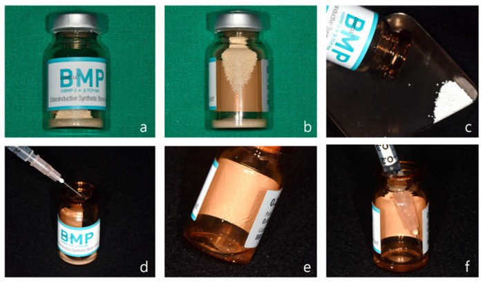
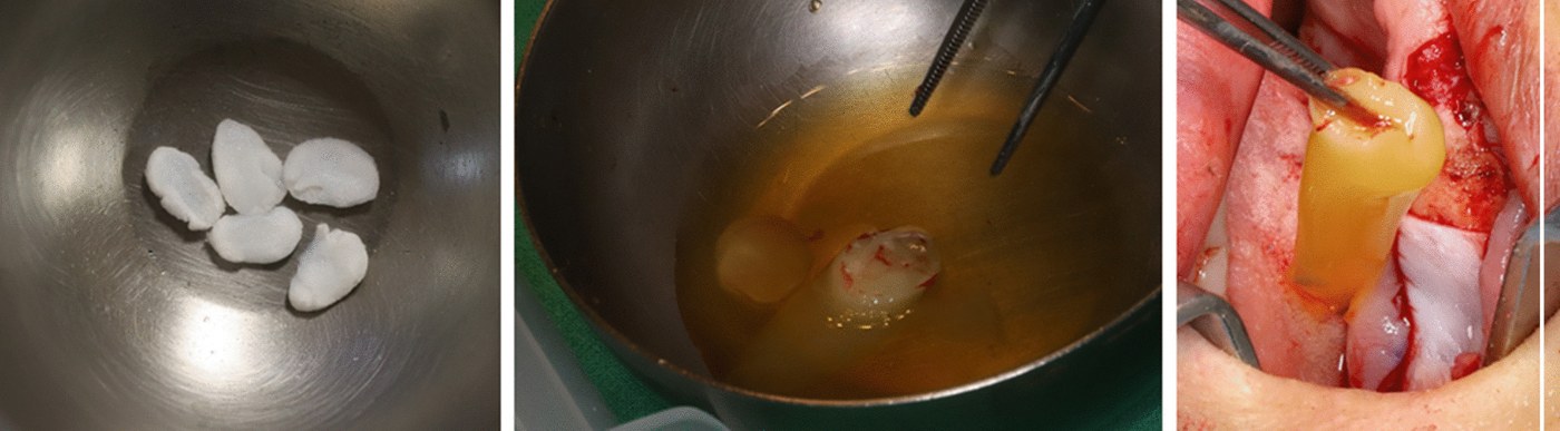
Examples of products.
| Product | Company | What it is | Status |
|---|---|---|---|
| Infuse Bone Graft (called InductOs in Europe) | Medtronic | rhBMP-2 on an absorbable collagen sponge | Approved for certain spinal fusions, open tibial fractures, and sinus and alveolar ridge augmentation |
| OP-1 Implant and OP-1 Putty | Stryker | rhBMP-7 with a collagen carrier | No longer marketed |
| Novosis | CGBio (Korea) | rhBMP-2 with hydroxyapatite or β-TCP granules | Available in parts of Asia |
| Augment | Wright Medical | A different growth factor: rhPDGF-BB with β-TCP | Foot and ankle fusion |
| GEM 21S | Osteohealth | rhPDGF-BB with β-TCP | Periodontal bone defects |
Evidence
| Use | Evidence |
|---|---|
| Open tibial fracture | Randomized trial of 450 patients. BMP added to nailing reduced the need for further surgery (Govender 2002). |
| Spinal fusion | The largest body of evidence. Two independent analyses of all the manufacturer's trial data found that rhBMP-2 increased fusion slightly, but gave no clinically important advantage over iliac crest graft (Simmonds 2013; Fu 2013). |
| Maxillary sinus floor augmentation | Randomized trial. BMP formed enough bone for dental implants (Boyne 2005). |
| Alveolar cleft in older patients | 21 skeletally mature patients. Bone fill 95% with BMP and 63% with iliac graft; complications 11% and 50% (Dickinson 2008). |
| Alveolar cleft in children | Used off-label in some centers with demineralized bone matrix. In 93 patients it failed more often in large clefts, as iliac graft did: failure was 65% above a cleft volume of about 0.9 cm³ and 14% below it (Roohani 2025). |
| Mandibular defects | Case series only (Herford and Boyne 2008). |
Risks and limitations
- BMP is only a signal. It has no cells and no strength. It works only in a bed with good blood supply and cells that can respond.
- Swelling. BMP causes a strong inflammatory reaction. This is dangerous near the airway. The FDA gave a warning about use in the anterior cervical spine in 2008. In a child with craniosynostosis, off-label use in the skull caused severe swelling of the scalp and face, which settled only after the implants were removed (Shah 2008).
- Bone in unwanted places (heterotopic bone), and early bone resorption around the implant.
- Cancer. At a high dose (40 mg) in spinal fusion, new cancers were more frequent with rhBMP-2 than with bone graft (Carragee 2013). In the pooled analyses the number of events was small. One found a raised risk and the other called the evidence inconclusive (Fu 2013; Simmonds 2013). BMP is therefore not placed in or near a tumor bed.
- Reporting. The early spine trials under-reported complications (Carragee 2011). The independent re-analysis confirmed this (Fu 2013).
- Approval. It is approved only for specific uses. Use in cleft and craniofacial surgery is off-label. It is avoided in young children, in pregnancy, and at sites of tumor or active infection (James 2016).
Position in plastic surgery. BMP can replace autograft in selected small, well-vascularized defects of the jaws in adults. It is not a substitute for vascularized bone in a large, irradiated or scarred defect. Most patients who have a mandibular resection have cancer, and this is a reason not to use it in them.
8B.2.3 Platelet concentrates
What they are
Platelets contain granules filled with growth factors: PDGF, TGF-β, VEGF and others. A platelet concentrate is made from the patient's own blood by centrifuge, so that a small volume contains many platelets.
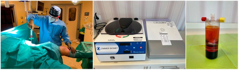
- Platelet-rich plasma (PRP) is plasma with a platelet count several times above normal. The commonly used working definition is about 1,000,000 platelets per microliter (Marx 2004). Blood is drawn with an anticoagulant. The PRP is a liquid and is activated at the time of use, for example with calcium.
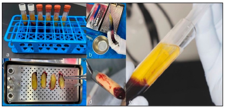
- Platelet-rich fibrin (PRF) is made without anticoagulant. The blood clots in the tube during spinning. The result is a fibrin clot that contains platelets and white cells and releases growth factors slowly. It can be pressed into a membrane (Choukroun 2006).
| Family | Form | White cells |
|---|---|---|
| Pure platelet-rich plasma (P-PRP) | Liquid; gel after activation | Few |
| Leukocyte- and platelet-rich plasma (L-PRP) | Liquid; gel after activation | Many |
| Pure platelet-rich fibrin (P-PRF) | Solid fibrin clot | Few |
| Leukocyte- and platelet-rich fibrin (L-PRF) | Solid fibrin clot | Many |
Classification of Dohan Ehrenfest and colleagues (2009).
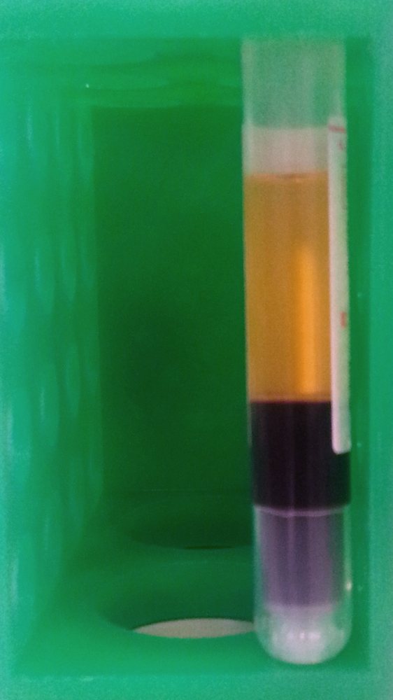
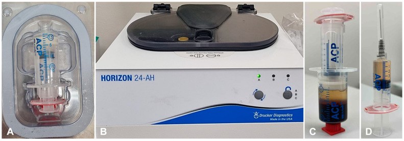
Examples of products
Each is a disposable kit used with its own centrifuge.
| System | Company | Product made |
|---|---|---|
| ACP double syringe | Arthrex | Platelet-rich plasma with few white cells |
| GPS III | Zimmer Biomet | Platelet-rich plasma with many white cells |
| SmartPrep | Harvest (Terumo) | Platelet-rich plasma; the same machine concentrates marrow |
| RegenKit | RegenLab | Platelet-rich plasma, separated by a gel in the tube |
| IntraSpin L-PRF | Intra-Lock | Platelet-rich fibrin clot and membrane |
Evidence
- Mandible. Marx (1998) added PRP to cancellous grafts in mandibular defects and found faster bone maturation on X-ray. This study started the clinical use of PRP in bone.
- Alveolar cleft. In 7 patients, iliac graft with PRP gave a higher volume of regenerated bone on CT than graft alone (Oyama 2004). The study was small.
- Long bones. A Cochrane review found only one small trial that met its criteria and concluded that the evidence was insufficient to support routine use (Griffin 2012).
How they work, and why the results differ
- The growth factors in platelets are supportive, not osteoinductive. PRP cannot make bone where no bone-forming cells are present.
- The products differ. Each system gives a different platelet count and white cell count, and each patient's platelets differ. A result from one system does not apply to another.
- The growth factors are released within hours to days, but a graft takes weeks to be incorporated.
Position in plastic surgery. PRP and PRF are safe, because they are the patient's own blood, and inexpensive. They may help soft tissue healing over a graft, and PRF can be used as a membrane to hold particulate graft. There is no good evidence that they increase bone union. They are optional additions to a graft, never the graft itself.
8B.2.4 Systemic drugs
Some drugs given for osteoporosis increase bone formation, and have been tested for fracture healing.
- Teriparatide is the active part of parathyroid hormone (amino acids 1 to 34). Given once daily by injection it stimulates osteoblasts.
- It is sold as Forteo (Forsteo in Europe) by Eli Lilly, for osteoporosis.
- In a randomized trial of 102 women with distal radius fractures, the median time to healing on X-ray was 9.1 weeks with placebo, 7.4 weeks with 20 µg and 8.8 weeks with 40 µg. The main hypothesis, that the higher dose would shorten healing, was not supported (Aspenberg 2010).
- It is not approved for fracture or graft healing. Its use for this purpose is off-label.
The opposite group of drugs matters more often in our practice. Bisphosphonates and denosumab suppress osteoclasts and bone turnover. In patients taking them, surgery on the jaw carries a risk of medication-related osteonecrosis of the jaw.
8B.3 Products that supply cells
8B.3.1 The mesenchymal stem cell
A mesenchymal stem cell (MSC), also called a mesenchymal stromal cell, is a cell that can divide and can become bone, cartilage or fat (Pittenger 1999). The accepted definition has three parts (Dominici 2006):
- it attaches to plastic in culture;
- it carries the surface markers CD105, CD73 and CD90, and lacks the blood cell markers CD45, CD34, CD14 and HLA-DR;
- it can become osteoblasts, chondrocytes and fat cells in the laboratory.
MSCs are found in marrow, periosteum, fat and other tissues. In the body they act in two ways: some become bone-forming cells, and many release signals that bring vessels and other cells.
8B.3.2 Bone marrow aspirate and concentrate
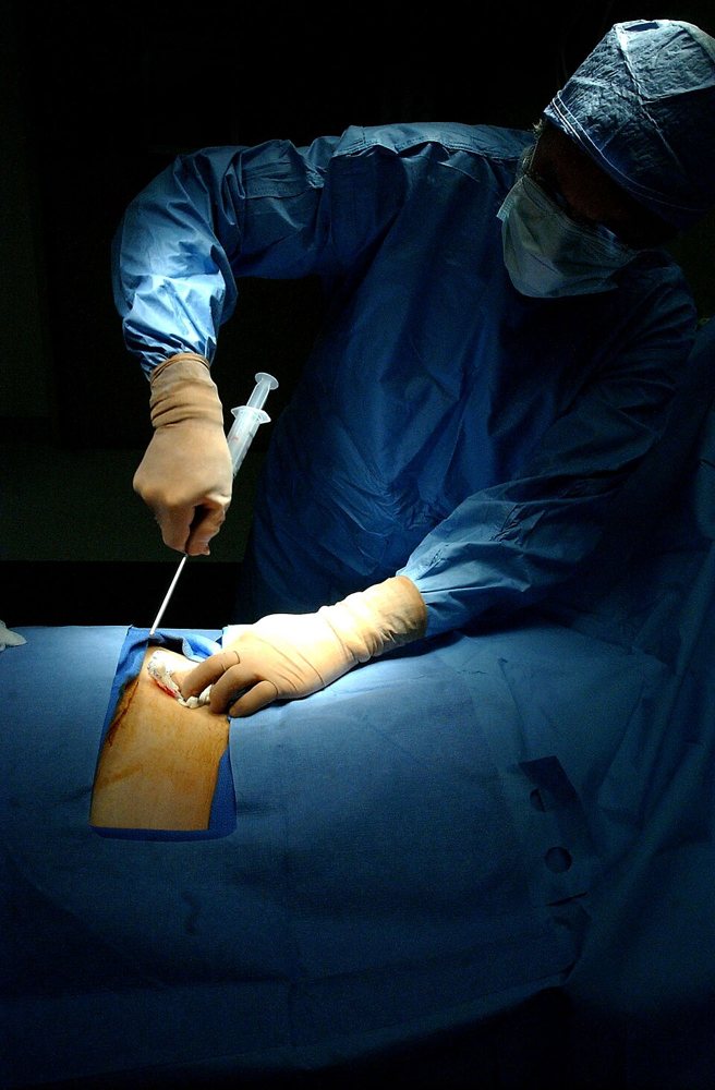
- Marrow is aspirated from the iliac crest with a needle. It can be used directly (bone marrow aspirate, BMA) or concentrated by centrifuge (bone marrow aspirate concentrate, BMAC).
- MSCs are rare. Only about 1 in 10,000 to 100,000 nucleated marrow cells is an MSC, and the number falls with age. In one study the mean was 36 bone-forming progenitor cells per million nucleated cells (Muschler 1997).
- Technique matters. Aspirating more than 2 mL from one site dilutes the marrow with blood. Four aspirates of 1 mL from different sites give almost twice as many progenitor cells as one aspirate of 4 mL (Muschler 1997).
- The dose matters. Hernigou (2005) injected concentrated marrow into 60 atrophic tibial nonunions. 53 united. The 7 that failed had received fewer progenitor cells.
- The limit. The number of stem cells varies greatly between patients and cannot be known at the time of injection.
- Marrow has no structure. It is combined with a scaffold, such as allograft or a ceramic, to make a "composite graft".
Examples of products
Marrow is aspirated with a marrow needle of the Jamshidi type and concentrated in a disposable kit: SmartPrep BMAC (Harvest, Terumo), Angel (Arthrex) and BioCUE (Zimmer Biomet) are examples.
8B.3.3 Adipose-derived stem cells
Fat is of special interest to plastic surgeons, because we harvest it routinely.
- In 2001 Zuk and colleagues showed that fat removed by liposuction contains cells that can become bone, cartilage, muscle and fat cells in the laboratory (Zuk 2001). These are called adipose-derived stem cells (ASCs).
- Fat gives many more stem cells per milliliter than marrow, and is easy to harvest.
- First clinical report in bone. A 7-year-old girl with large skull defects after trauma was treated with her own fat-derived cells, a small amount of iliac bone and fibrin glue (Lendeckel 2004).
- Case series. In 13 craniomaxillofacial bone defects (skull, frontal sinus, mandible, nasal septum), ASCs were placed on bioactive glass or β-tricalcium phosphate scaffolds, in some cases with rhBMP-2. The construct integrated in 10 of 13 (Sándor 2014).
- These cells were grown in a laboratory before use. This needs a licensed facility and regulatory approval, and it is not routine practice.
Position in plastic surgery. Marrow aspirate is a simple, low-risk way to add cells to a scaffold or allograft in a small defect with a good bed. Adipose-derived cells for bone are still experimental. Fat grafting as we do it for contour does not make bone.
8B.4 Combining scaffold, cells and signal: tissue engineering
Tissue engineering combines a scaffold, cells and a signal to build new bone. The three together are called the tissue engineering triad. With a blood supply and mechanical stability, they are the same requirements as the diamond concept.
Flowchart: What tissue-engineered bone needs
- Tissue-engineered bone
- Scaffoldceramic, mesh, printed polymer
- Cellsmarrow or fat-derived stem cells
- SignalBMP
- Blood supplythe main unsolved problem
Long bone. Quarto and colleagues (2001) reported the repair of large long bone defects with the patient's own marrow stromal cells, grown in the laboratory and placed on a porous hydroxyapatite scaffold.
Mandible: a case that used a flap as the bioreactor. Warnke (2004) reported one patient with a large mandibular defect:
- A titanium mesh cage was made in the shape of the missing mandible (scaffold).
- It was filled with bone mineral blocks, 7 mg of BMP-7 (signal) and 20 mL of the patient's bone marrow (cells).
- It was implanted in the latissimus dorsi muscle for 7 weeks (blood supply).
- It was then transferred to the jaw as a free flap. Bone scan showed living bone.
This case shows the four requirements used together. It also shows the idea of flap prefabrication: growing a construct inside a muscle so that it gains a blood supply and can then be moved as a flap. This is a plastic surgery contribution to the field.
However, these are single cases and small series. The standard treatment for a large mandibular defect is still the free fibula flap. The main limit of tissue-engineered bone is still blood supply: a construct larger than a few millimeters cannot survive on diffusion.
8B.5 Creating the vascular bed: the induced membrane (Masquelet) technique
All the products above assume a bed with a good blood supply. When the bed itself is what is missing, it must be created first. The induced membrane technique is a way to do this. It was described by Masquelet, a reconstructive surgeon, for large bone defects in limbs (Masquelet 2000). It is done in two stages.
- The defect. A large segmental defect, for example after debridement of an open fracture or osteomyelitis. Cancellous graft placed directly into such a defect is resorbed before vessels reach it.
- Stage 1. Debridement. The defect is filled with a bone cement (PMMA) spacer. The bone is stabilized. The wound is covered, usually with a flap.
- Wait 6 to 8 weeks. The body forms a membrane around the cement, as it does around any foreign body. This membrane has many vessels and produces growth factors (VEGF, TGF-β1, BMP-2) (Pelissier 2004).
- Stage 2. The membrane is opened and the spacer is removed. The space is filled with cancellous autograft and the membrane is closed. The membrane supplies the graft and prevents resorption. Masquelet's first report had 35 cases.
Flowchart: Induced membrane (Masquelet) technique
- Debridement
- Cement spacer + fixation + flap coverstage 1
- Wait 6 to 8 weeksmembrane forms
- Remove spacer, fill with cancellous autograftstage 2
- Union
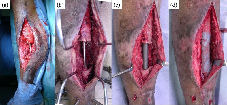
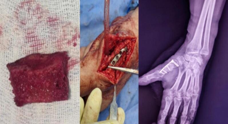
The cement. The spacer is made from ordinary PMMA bone cement, for example Palacos (Heraeus) or Simplex P (Stryker). Antibiotic powder is often added when the defect followed an infection.
Results.
- A systematic review found that infection persisted or the bone failed to unite in 18% of cases, which then needed further surgery (Morelli 2016).
- In the hand. In 11 severe hand and wrist injuries with bone loss treated by this technique as an emergency, 9 united, within a mean of four months (Flamans 2010).
The role of the plastic surgeon is the flap cover in stage 1. Without good soft tissue cover the technique cannot be done. In the hand, the plastic surgeon may do the whole procedure.
(Masquelet 2000; Masquelet and Begue 2010)
8B.6 Summary
- An orthobiologic supplies one part of what bone healing needs. It is an addition to stability, blood supply and a proper graft.
- BMP is the only osteoinductive product. It works, but at very high doses, with swelling, unwanted bone and a possible cancer risk. In craniofacial surgery its use is off-label.
- Platelet products supply supportive growth factors. They are safe, but have not been shown to improve bone union.
- Marrow supplies cells, in small and unpredictable numbers. The aspiration technique affects the yield.
- Fat is a rich source of stem cells, but their use for bone is experimental.
- The induced membrane technique creates a vascular bed for a large graft. It depends on good flap cover.
- Tissue-engineered bone is limited by blood supply. Vascularized bone remains the standard for large defects.
References
- Urist MR. Bone: formation by autoinduction. Science 1965;150:893–899.
- Govender S, Csimma C, Genant HK, et al. Recombinant human bone morphogenetic protein-2 for treatment of open tibial fractures. J Bone Joint Surg Am 2002;84:2123–2134.
- Friedlaender GE, Perry CR, Cole JD, Cook SD, et al. Osteogenic protein-1 (bone morphogenetic protein-7) in the treatment of tibial nonunions. J Bone Joint Surg Am 2001;83-A Suppl 1:S151–S158.
- Simmonds MC, Brown JV, Heirs MK, Higgins JP, et al. Safety and effectiveness of recombinant human bone morphogenetic protein-2 for spinal fusion: a meta-analysis of individual-participant data. Ann Intern Med 2013;158:877–889.
- Fu R, Selph S, McDonagh M, Peterson K, et al. Effectiveness and harms of recombinant human bone morphogenetic protein-2 in spine fusion: a systematic review and meta-analysis. Ann Intern Med 2013;158:890–902.
- Boyne PJ, Lilly LC, Marx RE, et al. De novo bone induction by rhBMP-2 in maxillary sinus floor augmentation. J Oral Maxillofac Surg 2005;63:1693–1707.
- Dickinson BP, Ashley RK, Wasson KL, et al. Reduced morbidity and improved healing with bone morphogenic protein-2 in older patients with alveolar cleft defects. Plast Reconstr Surg 2008;121:209–217.
- Roohani I, Youn S, Alfeerawi S, Shakoori P, et al. Failure rates based on alveolar cleft volume: analysis of the critical-size defect for alveolar bone grafting. Plast Reconstr Surg 2025;155:377e–386e.
- Herford AS, Boyne PJ. Reconstruction of mandibular continuity defects with bone morphogenetic protein-2. J Oral Maxillofac Surg 2008;66:616–624.
- Shah MM, Smyth MD, Woo AS. Adverse facial edema associated with off-label use of recombinant human bone morphogenetic protein-2 in cranial reconstruction for craniosynostosis. J Neurosurg Pediatr 2008;1:255–257.
- Carragee EJ, Hurwitz EL, Weiner BK. A critical review of recombinant human bone morphogenetic protein-2 trials in spinal surgery. Spine J 2011;11:471–491.
- Carragee EJ, Chu G, Rohatgi R, Hurwitz EL, et al. Cancer risk after use of recombinant bone morphogenetic protein-2 for spinal arthrodesis. J Bone Joint Surg Am 2013;95:1537–1545.
- James AW, LaChaud G, Shen J, et al. A review of the clinical side effects of bone morphogenetic protein-2. Tissue Eng Part B Rev 2016;22:284–297.
- Marx RE, Carlson ER, Eichstaedt RM, et al. Platelet-rich plasma: growth factor enhancement for bone grafts. Oral Surg Oral Med Oral Pathol Oral Radiol Endod 1998;85:638–646.
- Marx RE. Platelet-rich plasma: evidence to support its use. J Oral Maxillofac Surg 2004;62:489–496.
- Choukroun J, Diss A, Simonpieri A, Girard MO, et al. Platelet-rich fibrin (PRF): a second-generation platelet concentrate. Part IV: clinical effects on tissue healing. Oral Surg Oral Med Oral Pathol Oral Radiol Endod 2006;101:e56–e60.
- Dohan Ehrenfest DM, Rasmusson L, Albrektsson T. Classification of platelet concentrates: from pure platelet-rich plasma (P-PRP) to leucocyte- and platelet-rich fibrin (L-PRF). Trends Biotechnol 2009;27:158–167.
- Oyama T, Nishimoto S, Tsugawa T, Shimizu F. Efficacy of platelet-rich plasma in alveolar bone grafting. J Oral Maxillofac Surg 2004;62:555–558.
- Griffin XL, Wallace D, Parsons N, Costa ML. Platelet rich therapies for long bone healing in adults. Cochrane Database Syst Rev 2012;(7):CD009496.
- Pittenger MF, Mackay AM, Beck SC, Jaiswal RK, et al. Multilineage potential of adult human mesenchymal stem cells. Science 1999;284:143–147.
- Dominici M, Le Blanc K, Mueller I, Slaper-Cortenbach I, et al. Minimal criteria for defining multipotent mesenchymal stromal cells. The International Society for Cellular Therapy position statement. Cytotherapy 2006;8:315–317.
- Muschler GF, Boehm C, Easley K. Aspiration to obtain osteoblast progenitor cells from human bone marrow: the influence of aspiration volume. J Bone Joint Surg Am 1997;79:1699–1709.
- Hernigou P, Poignard A, Beaujean F, Rouard H. Percutaneous autologous bone-marrow grafting for nonunions. J Bone Joint Surg Am 2005;87:1430–1437.
- Zuk PA, Zhu M, Mizuno H, Huang J, et al. Multilineage cells from human adipose tissue: implications for cell-based therapies. Tissue Eng 2001;7:211–228.
- Lendeckel S, Jödicke A, Christophis P, Heidinger K, et al. Autologous stem cells (adipose) and fibrin glue used to treat widespread traumatic calvarial defects: case report. J Craniomaxillofac Surg 2004;32:370–373.
- Sándor GK, Numminen J, Wolff J, Thesleff T, et al. Adipose stem cells used to reconstruct 13 cases with cranio-maxillofacial hard-tissue defects. Stem Cells Transl Med 2014;3:530–540.
- Masquelet AC, Fitoussi F, Begue T, Muller GP. Reconstruction of the long bones by the induced membrane and spongy autograft. Ann Chir Plast Esthet 2000;45:346–353.
- Pelissier P, Masquelet AC, Bareille R, et al. Induced membranes secrete growth factors including vascular and osteoinductive factors. J Orthop Res 2004;22:73–79.
- Masquelet AC, Begue T. The concept of induced membrane for reconstruction of long bone defects. Orthop Clin North Am 2010;41:27–37.
- Morelli I, Drago L, George DA, Gallazzi E, et al. Masquelet technique: myth or reality? A systematic review and meta-analysis. Injury 2016;47 Suppl 6:S68–S76.
- Flamans B, Pauchot J, Petite H, Blanchet N, et al. Use of the induced membrane technique for the treatment of bone defects in the hand or wrist, in emergency. Chir Main 2010;29:307–314.
- Aspenberg P, Genant HK, Johansson T, Nino AJ, et al. Teriparatide for acceleration of fracture repair in humans: a prospective, randomized, double-blind study of 102 postmenopausal women with distal radial fractures. J Bone Miner Res 2010;25:404–414.
- Quarto R, Mastrogiacomo M, Cancedda R, Kutepov SM, et al. Repair of large bone defects with the use of autologous bone marrow stromal cells. N Engl J Med 2001;344:385–386.
- Warnke PH, Springer ING, Wiltfang J, et al. Growth and transplantation of a custom vascularised bone graft in a man. Lancet 2004;364:766–770.