11.1 Introduction
The patient's own bone has two disadvantages: donor site morbidity and limited amount. This chapter describes bone taken from another source.
| Term | Source |
|---|---|
| Autograft | The same patient |
| Allograft | Another human (living or deceased donor) |
| Xenograft | Another species (usually bovine) |
The main point of this chapter: allograft and xenograft have no living cells. They are not osteogenic. They act mainly as a scaffold (osteoconduction). Some have a weak osteoinductive effect.
Where we use them in plastic surgery:
- Large structural allograft combined with a free fibula in limb reconstruction
- Demineralized bone matrix to fill small gaps in craniofacial surgery
- Bovine bone for sinus floor and alveolar ridge augmentation before dental implants
- Allograft chips mixed with autograft when there is not enough autograft
Flowchart: Bone grafts by source
- Bone graft materials
- Autograftsame patient
- Has living cells
- Allograftanother human
- Fresh-frozenstructural
- Freeze-driedchips, small grafts
- Demineralized (DBM)putty; weak osteoinduction
- Xenograftbovine
- Bone mineral onlyslow to resorb
- Alloplastsynthetic
- No biological activity
- Autograftsame patient
11.2 Forms of allograft
- Cancellous chips: to fill contained defects.
- Corticocancellous blocks.
- Cortical struts and whole bone segments (massive allograft): for structural support.
- Demineralized bone matrix (DBM): powder, putty or gel.
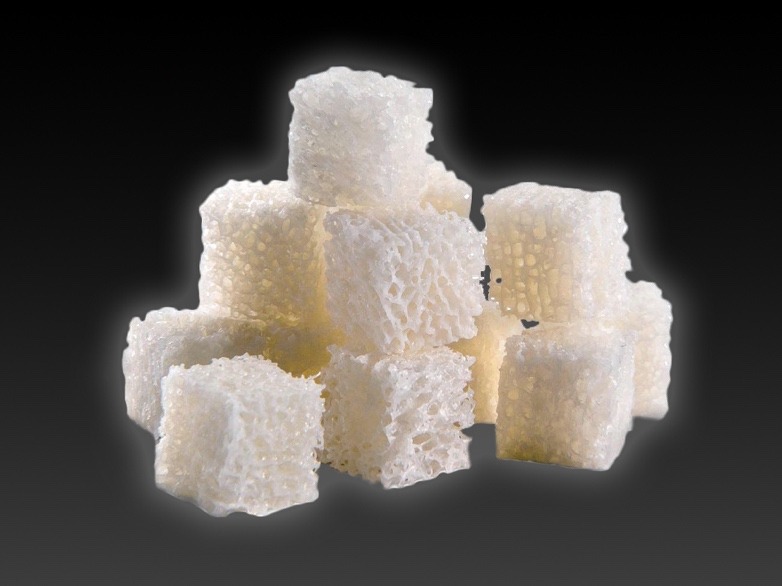
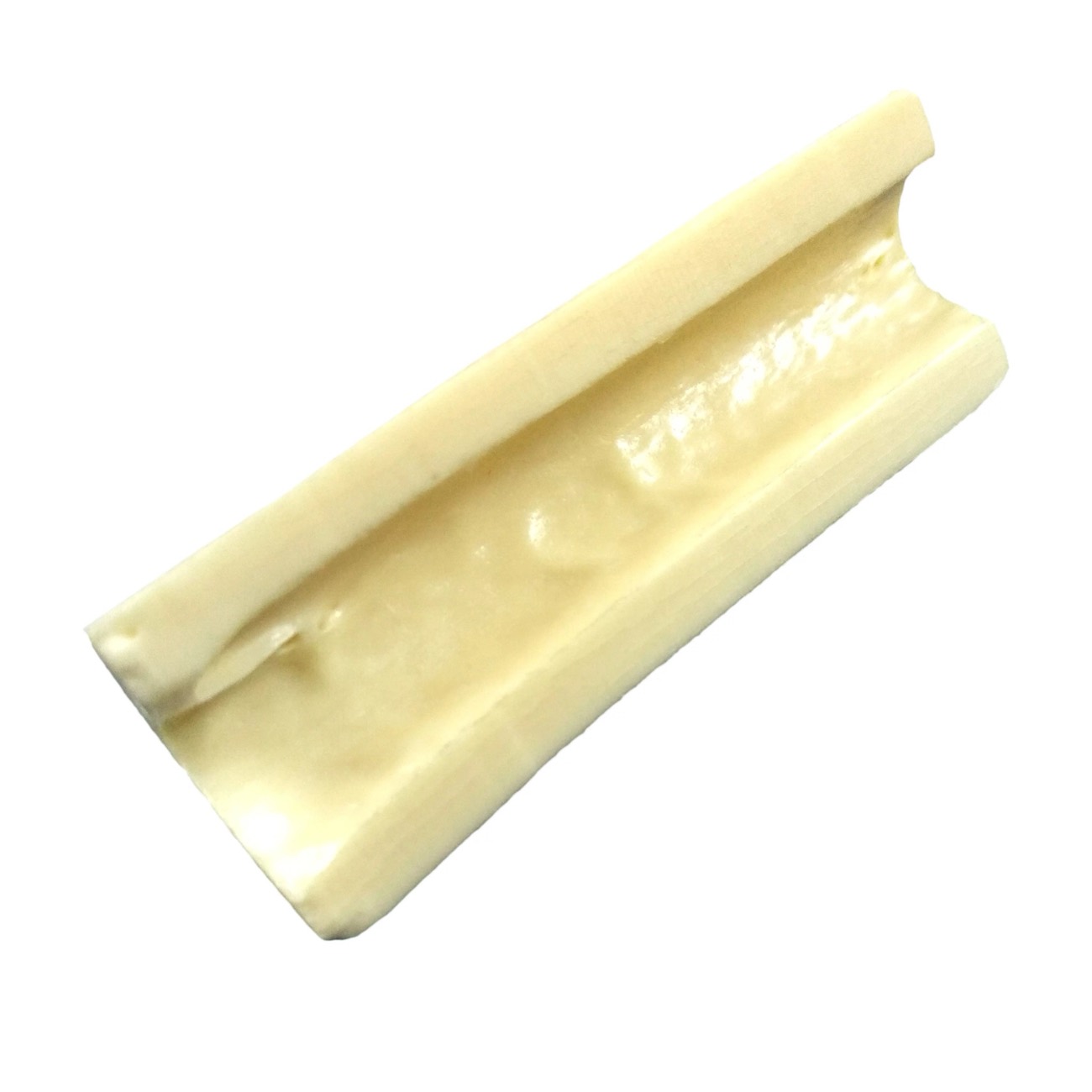
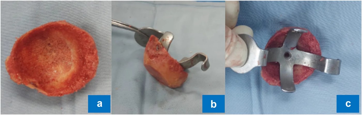
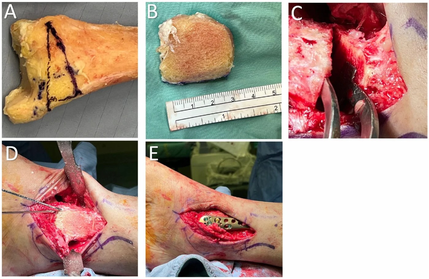
Where allograft comes from. Allograft bone is supplied by tissue banks, not by ordinary manufacturers. In Malaysia the USM Tissue Bank supplies processed bone. Large tissue banks abroad include MTF Biologics, LifeNet Health and AlloSource in the United States. Some processed allografts carry a brand name:
| Product | What it is | Supplier |
|---|---|---|
| Puros | Solvent-preserved cancellous or cortical allograft, in particles and blocks. It is processed by the Tutoplast method. | Zimmer Biomet |
| Osteocel | Cancellous allograft that is frozen in a way that keeps some donor cells alive | NuVasive |
| Trinity Evolution | The same idea: allograft with living donor cells | Orthofix, with MTF |
11.3 Processing of allograft
Allograft must be processed to make it safe and to allow storage. Each processing step reduces the risk of infection and rejection, but also reduces the strength or the biological activity of the graft.
| Type | Storage | Strength | Use |
|---|---|---|---|
| Fresh-frozen | −70 °C or below | Preserved | Structural (massive) allograft |
| Freeze-dried | Room temperature, for years | Reduced; brittle until rehydrated | Chips and small grafts for filling |
| Gamma-irradiated | (a sterilization step added to the above) | Reduced further, depending on dose | Standard dose is about 25 kGy |
| Demineralized | Room temperature | None | Gap filling |
Flowchart: Processing of allograft
- Donor bone
- Clean: remove soft tissue and marrow
- Freeze (−70 °C) or freeze-dry or demineralize
- Gamma irradiationabout 25 kGy
- Store and label
11.3.1 Details of processing
- Fresh-frozen: stored at −70 to −80 °C, for up to about 5 years. Freezing kills the cells and reduces immunogenicity. Mechanical properties are almost unchanged.
- Freeze-dried (lyophilized): the bone is frozen and the water is removed under vacuum to a residual moisture of a few percent. It is stored at room temperature. It must be rehydrated before use; dry bone is brittle, and bending and torsional strength are reduced.
- Gamma irradiation: 25 kGy is the conventional dose for a sterility assurance level of 10−6. Radiation breaks collagen chains by direct effect and by free radicals. The loss of strength is dose-dependent, and is larger for bending and fatigue than for compression.
- Demineralization: hydrochloric acid (about 0.5 to 0.6 N) removes the mineral.
- Ethylene oxide is no longer favored, because residues reduce osteoinduction and can cause inflammation.
11.4 Incorporation of allograft
Allograft incorporates in the same way as non-vascularized cortical autograft (creeping substitution), but more slowly and less completely.
- Enneking and Campanacci (2001) examined massive allografts removed from patients years after implantation. The allograft had united with the host bone at its ends. But new bone had replaced only the ends and a thin surface layer. Most of the graft was still dead bone.
- Dead bone cannot repair small cracks. So allografts can fracture late, and these fractures do not heal.
Results of massive allograft for bone tumors (Mankin 1996):
- Infection in about 10%, mostly in the first year.
- Fracture in about 19%, mostly by the third year.
- About 75% were retained and successful in the long term.
This is why a vascularized fibula is combined with the allograft (Capanna technique). The living fibula helps union and reduces these failures.
11.5 Demineralized bone matrix (DBM)
DBM is allograft bone treated with acid to remove the mineral. What remains is collagen and bone proteins, including small amounts of BMP. Removing the mineral exposes these proteins, so DBM is osteoinductive in principle. It has no strength.
- Urist (1965) discovered osteoinduction using demineralized bone.
- Mulliken and Glowacki (1980, 1981), plastic surgeons in Boston, were the first to use demineralized bone in patients with craniofacial defects.
- USM (Ali, Halim et al. 2005): a 7-year-old girl with an orbito-frontal bone defect was reconstructed with a vascularized outer-table calvarial bone graft on the superficial temporal vessels. The gaps around it were filled with DBM putty.
Commercial DBM is mixed with a carrier so that it can be handled: glycerol, hyaluronic acid, gelatin, collagen or calcium sulfate. The carrier may make up most of the volume. Products differ in particle size, carrier and sterilization method, and all of these affect osteoinductive activity.
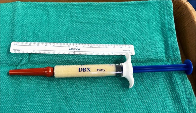
Limitation. The amount of BMP in commercial DBM is very small and not consistent. Bae (2006) measured only nanograms of BMP per gram of DBM, and found that the amount differed more between batches of the same product than between different products. (For comparison, an rhBMP-2 product contains milligrams.)
DBM is best used as a filler around living bone or autograft. It should not be used alone to bridge a defect.
Examples of DBM products. DBM is a powder. To make it usable it is mixed with a carrier, and the carrier differs between products.
| Product | Carrier and form | Supplier |
|---|---|---|
| Grafton | Glycerol; gel, putty, flexible sheets | Medtronic (first made by Osteotech) |
| DBX | Sodium hyaluronate; putty and paste | MTF Biologics, distributed by DePuy Synthes |
| Allomatrix | Calcium sulfate; putty that is mixed in the operating room | Wright Medical |
The amount of BMP differs between products and between batches of the same product. Two DBM products cannot be assumed to be equal.
11.6 Safety of allograft
Patients often ask two questions about allograft.
Will it be rejected?
- Bone allograft is not tissue-matched, and no immunosuppression is given.
- The antigens are on the donor cells and marrow. Processing (washing, freezing, freeze-drying) removes most of them.
- The remaining immune reaction slows incorporation, but it does not cause clinical rejection.
- The immune response is mainly cell-mediated, against MHC class I and II antigens on donor cells (Friedlaender 1983).
- Fresh allograft is the most immunogenic, frozen is less, freeze-dried is least.
- Histologically the response appears as a delay in revascularization and a fibrous layer around the graft.
Can it transmit infection?
The risk is very low but not zero. Among tissue donors in the United States, the estimated chance that a donor has an infection that is missed by antibody tests was (Zou 2004):
| Virus | Estimated risk per donor |
|---|---|
| Hepatitis B | 1 in 34,000 |
| Hepatitis C | 1 in 42,000 |
| HIV | 1 in 55,000 |
The risk to the recipient is much lower than this, because of donor history screening, more sensitive nucleic acid tests, processing and irradiation. With proper screening, the risk of HIV from bone allograft was estimated at less than 1 in a million (Buck 1989).
11.7 Xenograft
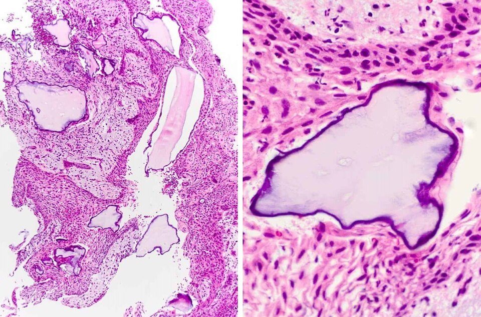
The xenograft in common use is deproteinized bovine bone mineral. All protein and cells are removed, leaving only the mineral structure of cancellous bone.
- It is osteoconductive only.
- It resorbs very slowly, over years. This is useful where volume must be maintained: maxillary sinus floor augmentation and alveolar ridge augmentation.
- It is not suitable where the graft must be fully replaced by normal bone, such as an alveolar cleft where a tooth must erupt.
Examples of xenograft products.
| Product | What it is | Supplier |
|---|---|---|
| Bio-Oss | Deproteinized bovine bone mineral; granules and blocks. The most studied product. | Geistlich |
| Endobon | Bovine hydroxyapatite, heated at high temperature | Zimmer Biomet |
| cerabone | Bovine bone mineral, heated at high temperature | botiss |
11.8 Summary
| Need | Material |
|---|---|
| Graft that is fully replaced by bone | Cancellous autograft (allograft chips can be added to increase volume) |
| Volume that is maintained | Bovine bone mineral |
| Structural support | Fresh-frozen structural allograft, preferably with vascularized bone |
| Filling small gaps | DBM |
References
- Nguyen H, Morgan DAF, Forwood MR. Sterilization of allograft bone: effects of gamma irradiation on allograft biology and biomechanics. Cell Tissue Bank 2007;8:93–105.
- Nguyen H, Morgan DAF, Forwood MR. Sterilization of allograft bone: is 25 kGy the gold standard for gamma irradiation? Cell Tissue Bank 2007;8:81–91.
- Enneking WF, Campanacci DA. Retrieved human allografts: a clinicopathological study. J Bone Joint Surg Am 2001;83:971–986.
- Mankin HJ, Gebhardt MC, Jennings LC, Springfield DS, Tomford WW. Long-term results of allograft replacement in the management of bone tumors. Clin Orthop Relat Res 1996;324:86–97.
- Capanna R, Campanacci DA, Belot N, et al. A new reconstructive technique for intercalary defects of long bones. Orthop Clin North Am 2007;38:51–60.
- Urist MR. Bone: formation by autoinduction. Science 1965;150:893–899.
- Mulliken JB, Glowacki J. Induced osteogenesis for repair and construction in the craniofacial region. Plast Reconstr Surg 1980;65:553–560.
- Mulliken JB, Glowacki J, Kaban LB, Folkman J, et al. Use of demineralized allogeneic bone implants for the correction of maxillocraniofacial deformities. Ann Surg 1981;194:366–372.
- Glowacki J, Kaban LB, Murray JE, Folkman J, et al. Application of the biological principle of induced osteogenesis for craniofacial defects. Lancet 1981;1:959–962.
- Ali F, Halim AS, Najihah SZ, Ibrahim M, Abdullah J. Combination of vascularized outer-table calvarial bone graft based on the superficial temporal vessels and allomatrix for the repair of an orbito-frontal blow-out fracture in a child. J Craniomaxillofac Surg 2005;33:326–330.
- Bae HW, Zhao L, Kanim LEA, et al. Intervariability and intravariability of bone morphogenetic proteins in commercially available demineralized bone matrix products. Spine 2006;31:1299–1306.
- Zou S, Dodd RY, Stramer SL, Strong DM, et al. Probability of viremia with HBV, HCV, HIV, and HTLV among tissue donors in the United States. N Engl J Med 2004;351:751–759.
- Buck BE, Malinin TI, Brown MD. Bone transplantation and human immunodeficiency virus: an estimate of risk of acquired immunodeficiency syndrome. Clin Orthop Relat Res 1989;240:129–136.
- Friedlaender GE. Immune responses to osteochondral allografts: current knowledge and future directions. Clin Orthop Relat Res 1983;174:58–68.