5.1 Introduction
Bone is formed in two ways. In intramembranous ossification, cells in a sheet of connective tissue (membrane) become osteoblasts and make bone directly. In endochondral ossification, a model of the bone is first made in cartilage, and the cartilage is then replaced by bone.
Both matter to the plastic surgeon. The skull vault and most of the face are made by the first route; the limbs, the skull base and the mandibular condyle by the second. A fracture heals by using both routes again. And in a child, the surgeon must protect not only the bone but its ability to grow: the physis in a limb, the sutures in the skull, and the growth sites of the face.
Flowchart: Two ways of forming bone
- Bone formation (ossification)
- Intramembranous
- Mesenchymal cells become osteoblasts directlyno cartilage stage
- Skull vault, most facial bones, body of the mandible, clavicle (in part)
- Mesenchymal cells become osteoblasts directlyno cartilage stage
- Endochondral
- A cartilage model is made firstvessels invade it, and bone replaces it
- Long bones, vertebrae, ribs, skull base, mandibular condyle
- A cartilage model is made firstvessels invade it, and bone replaces it
- Intramembranous
5.2 Embryonic origin of the skeleton
Before bone forms, the cells that will make it come from three sources in the embryo:
| Source | What it forms |
|---|---|
| Cranial neural crest | Most of the facial skeleton (nasal bones, maxilla, zygoma, mandible) and the frontal bone |
| Paraxial mesoderm | The parietal bones and parts of the skull base and occipital bone; the vertebrae and ribs |
| Lateral plate mesoderm | The bones of the limbs, the shoulder and pelvic girdles, and the sternum |
In the mouse skull, the frontal bone comes from neural crest and the parietal bone from mesoderm. The coronal suture forms exactly where the two tissues meet (Jiang 2002). This boundary helps explain why the coronal suture is so often involved in syndromic craniosynostosis.
Embryonic origin and route of ossification are different things. The mandible comes from neural crest, but its body forms by intramembranous ossification and its condyle by endochondral ossification.
5.3 Intramembranous ossification
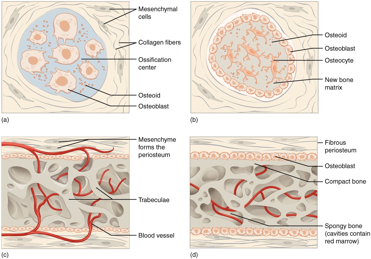
- Mesenchymal cells gather in a vascular membrane and become osteoblasts. This needs RUNX2.
- The osteoblasts lay down osteoid, which mineralizes. Some are trapped and become osteocytes.
- Trabeculae of woven bone form and join around blood vessels.
- The mesenchyme on the surface becomes the periosteum.
- The woven bone is remodeled into lamellar bone: compact bone at the surfaces (the outer and inner tables of the skull) and cancellous bone between (the diploë).
5.4 Endochondral ossification
- Mesenchymal cells gather and become cartilage cells (chondrocytes), under the control of the transcription factor SOX9. They make a small cartilage model of the bone.
- The model grows. Cells in its center enlarge (hypertrophy) and the matrix around them calcifies.
- The perichondrium around the middle of the model becomes periosteum and makes a bone collar by intramembranous ossification.
- Blood vessels invade the calcified center, bringing osteoclasts and osteoprogenitors. This is the primary ossification center, in the future diaphysis.
- Osteoblasts lay bone on the remaining struts of calcified cartilage. This mixed tissue is the primary spongiosa.
- The primary spongiosa is remodeled into trabecular bone, and a marrow cavity forms.
- Later, secondary ossification centers appear in the epiphyses. Cartilage remains in two places: the joint surface (articular cartilage) and the plate between epiphysis and metaphysis (the physis).
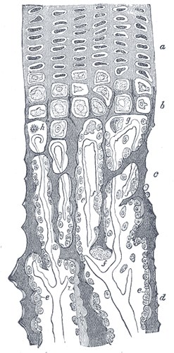
Calcified cartilage is only a temporary scaffold. It is mineralized, but it is not bone.
The hypertrophic cartilage cells were long thought simply to die. Lineage tracing in mice has shown that some of them survive and become osteoblasts and osteocytes (Yang 2014). Endochondral bone is therefore made partly by cells that were once cartilage cells.
5.5 The physis (growth plate)
Long bones grow in length at the physis. It is a disc of cartilage in which cells are made, line up, enlarge and are replaced by bone, in a fixed order from the epiphysis toward the metaphysis.
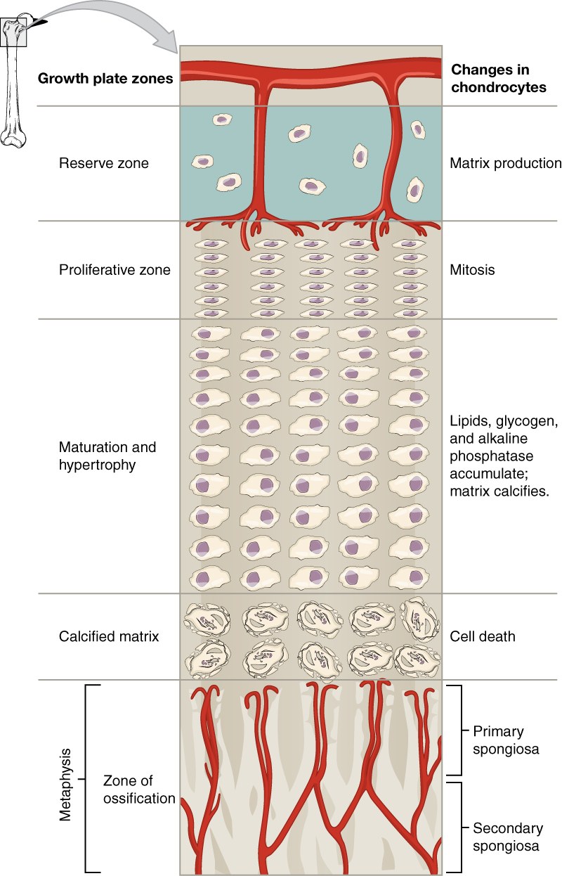
| Zone | What happens | Note |
|---|---|---|
| Reserve (resting) | Small, scattered cells | Contains the stem cells of the growth plate. In mice, these cells make PTHrP and supply the columns for the whole of growth (Mizuhashi 2018). |
| Proliferative | Cells divide and stack in columns | The main source of new cells |
| Hypertrophic | Cells enlarge up to ten times; type X collagen is made | Cell enlargement produces most of the increase in length. The weakest zone; most physeal fractures pass through it. |
| Calcification | Matrix calcifies; many cells die | Calcified struts remain |
| Ossification | Metaphyseal vessels invade; bone is laid on the struts | Forms the primary spongiosa |
Around the edge of the physis there are two structures. The groove of Ranvier contains cells that add to the width of the physis. The perichondrial ring of LaCroix is a fibrous band that holds the physis together and is continuous with the periosteum.
5.5.1 How growth is controlled
The speed of growth is set by a feedback loop between two signals (Vortkamp 1996; Kronenberg 2003):
Flowchart: The IHH and PTHrP feedback loop
- Cells about to enlarge make Indian hedgehog (IHH)
- IHH reaches the ends of the boneand stimulates the proliferating cells
- Cells near the ends make PTHrPparathyroid hormone-related protein
- PTHrP keeps cells dividingand delays their enlargement
This loop keeps the columns at a constant length. Other controls:
| Regulator | Effect | Example of disease |
|---|---|---|
| FGFR3 | Slows proliferation and enlargement | An activating mutation causes achondroplasia (Shiang 1994): short limbs, a small skull base and midface hypoplasia |
| Growth hormone and IGF-1 | Drive proliferation and enlargement | Deficiency causes short stature |
| Thyroid hormone | Needed for maturation of the plate | Deficiency delays bone age |
| Estrogen (in both sexes) | Speeds maturation; closes the physis at the end of puberty | Early puberty causes early closure |
| Glucocorticoids in excess | Suppress chondrocytes | Children on long-term steroids grow less |
5.5.2 Closure of the physis and injury
Toward the end of puberty, the physis thins and is replaced by bone. What remains is the epiphyseal line.
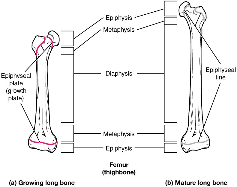
If part of the physis is injured in a child, bone can form across it. This physeal bar tethers growth on one side, causing angular deformity, or stops growth, causing shortening. A physeal fracture can unite normally and still cause a deformity a year later. Children with these injuries are followed until growth ends.
5.5.3 Growth in width
Long bones grow in width by appositional growth: osteoblasts under the periosteum add bone to the outer surface, while osteoclasts remove bone from the inner surface, enlarging the marrow cavity. This changes the size and shape of the bone without a remodeling cycle, a process called modeling.
5.6 Growth of the skull
5.6.1 Sutures and fontanelles
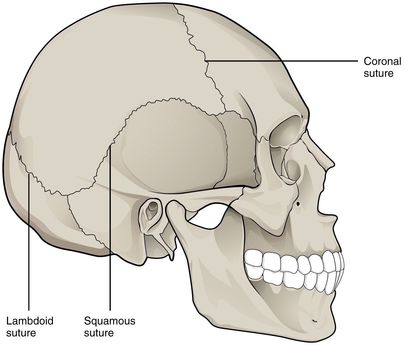
The bones of the skull vault are separated by sutures, bands of fibrous tissue between the bone edges. Where several bones meet, the gaps are larger and are called fontanelles. The anterior fontanelle usually closes by about 18 to 24 months.
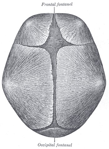
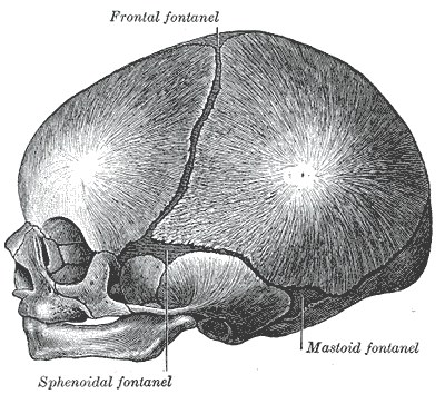
A suture is not a passive seam. It is the growth site of the skull vault. As the brain grows, it pushes the bones apart, and new bone is added at the edges of the bones on either side of the suture. The suture also contains stem cells for the bones of the skull: in mice, these cells are found in the suture, they repair injury, and removing them causes the suture to fuse (Zhao 2015).
5.6.2 Craniosynostosis
Craniosynostosis is the early fusion of one or more sutures. Growth stops across the fused suture, and the skull grows more at the open sutures instead. The shape of the head therefore depends on which suture has fused:
| Suture fused | Head shape |
|---|---|
| Sagittal | Scaphocephaly: long and narrow |
| One coronal | Anterior plagiocephaly: flat forehead on one side |
| Both coronal | Brachycephaly: short and wide |
| Metopic | Trigonocephaly: triangular forehead |
| One lambdoid | Posterior plagiocephaly |
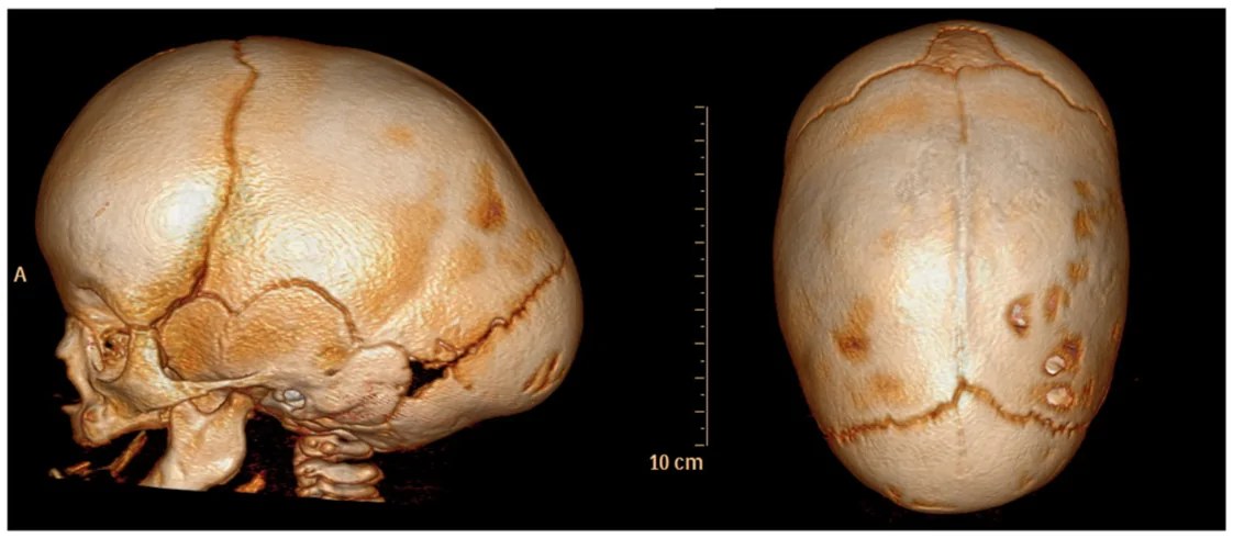
Most craniosynostosis is non-syndromic. Syndromic forms (Apert, Crouzon, Pfeiffer, Muenke, Saethre-Chotzen) are caused by mutations in genes that control the suture, mainly FGFR2, FGFR3 and TWIST1 (Morriss-Kay and Wilkie 2005).
5.6.3 Growth of the skull base and face
| Region | How it grows | Why it matters |
|---|---|---|
| Skull vault | Intramembranous; at the sutures | Craniosynostosis; vault remodeling |
| Skull base | Endochondral; at cartilage joints called synchondroses (for example the spheno-occipital synchondrosis) | Short skull base and midface retrusion in achondroplasia and in Apert and Crouzon syndromes |
| Maxilla | Intramembranous; at its sutures, by surface remodeling, and by being carried forward with the skull base | Scar from early palate repair can restrict maxillary growth in cleft patients |
| Mandible | Body and ramus intramembranous; the condyle has a cartilage layer (secondary cartilage) that grows endochondrally | Condylar injury in a child can cause asymmetry and restricted growth; mandibular distraction lengthens the bone |
The face does not grow only by its own program. According to the functional matrix theory, the bones grow in response to the soft tissues and spaces around them: the brain, the eyes, the airway, the muscles of chewing (Moss and Salentijn 1969). This explains why a small orbit follows loss of an eye in childhood, and why the face can grow abnormally when the airway or the muscles are abnormal.
5.7 Development and repair
Fracture healing uses the same two routes of bone formation:
- Where the fracture is stable and well vascularized, mainly under the periosteum near the fracture, bone forms directly, as in intramembranous ossification.
- Where there is movement and low oxygen, in the gap between the bone ends, cartilage forms first and is then replaced by bone, as in endochondral ossification.
Secondary (indirect) fracture healing therefore contains both routes. In distraction osteogenesis, if the frame is stable and the rate is right, bone in the gap forms mainly by the direct route. If there is too much movement, cartilage appears.
Repair copies development, but it is not a repeat of it. It happens in an adult bone, in the presence of injury, inflammation and an implant.
5.8 Summary
- Bone forms either directly from membrane (intramembranous) or through a cartilage model (endochondral).
- The skull vault and most of the face are intramembranous. The limbs, skull base and mandibular condyle are endochondral.
- Embryonic origin (neural crest or mesoderm) is separate from the route of ossification. The coronal suture lies at the neural crest–mesoderm boundary.
- The physis has reserve, proliferative, hypertrophic, calcification and ossification zones. Growth is controlled by the IHH–PTHrP loop, FGFR3 and hormones.
- A physeal bar can cause deformity after a fracture has united.
- Sutures are growth sites containing stem cells. Early fusion causes craniosynostosis, with a head shape that depends on the suture.
- The face grows at sutures, synchondroses and the condylar cartilage, and in response to the soft tissues around it.
- Fracture healing uses both routes of bone formation again.
References
- Jiang X, Iseki S, Maxson RE, Sucov HM, Morriss-Kay GM. Tissue origins and interactions in the mammalian skull vault. Dev Biol 2002;241:106–116.
- Yang G, Zhu L, Hou N, Lan Y, Wu XM, Zhou B, et al. Osteogenic fate of hypertrophic chondrocytes. Cell Res 2014;24:1266–1269.
- Mizuhashi K, Ono W, Matsushita Y, Sakagami N, Takahashi A, Saunders TL, et al. Resting zone of the growth plate houses a unique class of skeletal stem cells. Nature 2018;563:254–258.
- Vortkamp A, Lee K, Lanske B, Segre GV, Kronenberg HM, Tabin CJ. Regulation of rate of cartilage differentiation by Indian hedgehog and PTH-related protein. Science 1996;273:613–622.
- Kronenberg HM. Developmental regulation of the growth plate. Nature 2003;423:332–336.
- Shiang R, Thompson LM, Zhu YZ, Church DM, Fielder TJ, Bocian M, et al. Mutations in the transmembrane domain of FGFR3 cause the most common genetic form of dwarfism, achondroplasia. Cell 1994;78:335–342.
- Zhao H, Feng J, Ho TV, Grimes W, Urata M, Chai Y. The suture provides a niche for mesenchymal stem cells of craniofacial bones. Nat Cell Biol 2015;17:386–396.
- Morriss-Kay GM, Wilkie AO. Growth of the normal skull vault and its alteration in craniosynostosis: insights from human genetics and experimental studies. J Anat 2005;207:637–653.
- Moss ML, Salentijn L. The primary role of functional matrices in facial growth. Am J Orthod 1969;55:566–577.